1PGR | pdb_00001pgr

2:2 COMPLEX OF G-CSF WITH ITS RECEPTOR


Experimental Data Snapshot

  • Method: X-RAY DIFFRACTION
  • Resolution: 3.50 Å
  • R-Value Free: 
    0.281 (DCC) 
  • R-Value Work: 
    0.317 (Depositor), 0.290 (DCC) 

Starting Model: experimental
View more details

wwPDB Validation 3D Report Full Report

Validation slider image for 1PGR

This is version 1.4 of the entry. See complete history

Literature

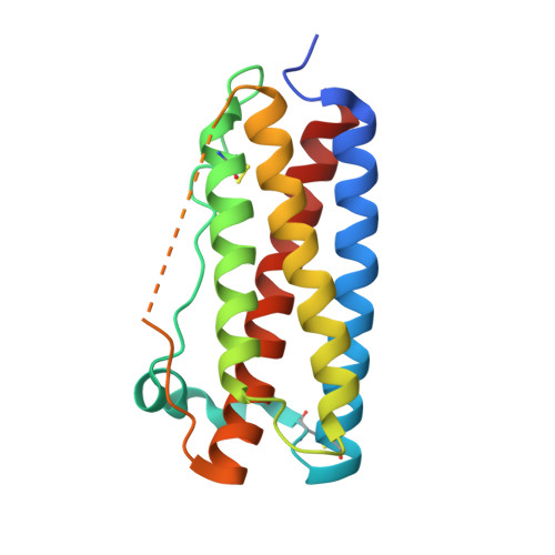
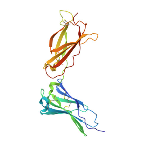
Atomic structure of the GCSF-receptor complex showing a new cytokine-receptor recognition scheme.

Aritomi, M.Kunishima, N.Okamoto, T.Kuroki, R.Ota, Y.Morikawa, K.

(1999) Nature 401: 713-717

  • DOI: https://doi.org/10.1038/44394
  • Primary Citation Related Structures: 
    1CD9, 1PGR

  • PubMed Abstract: 

    Granulocyte colony-stimulating factor (GCSF) is the principal growth factor regulating the maturation, proliferation and differentiation of the precursor cells of neutrophilic granulocytes and is used to treat neutropenia. GCSF is a member of the long-chain subtype of the class 1 cytokine superfamily, which includes growth hormone, erythropoietin, interleukin 6 and oncostatin M. Here we have determined the crystal structure of GCSF complexed to the BN-BC domains, the principal ligand-binding region of the GCSF receptor (GCSFR). The two receptor domains form a complex in a 2:2 ratio with the ligand, with a non-crystallographic pseudo-twofold axis through primarily the interdomain region and secondarily the BC domain. This structural view of a gp130-type receptor-ligand complex presents a new molecular basis for cytokine-receptor recognition.


  • Organizational Affiliation
    • Biomolecular Engineering Research Institute, Suita-city, Osaka, Japan.

Macromolecule Content 

  • Total Structure Weight: 173.11 kDa 
  • Atom Count: 11,424 
  • Modeled Residue Count: 1,471 
  • Deposited Residue Count: 1,560 
  • Unique protein chains: 2

Macromolecules

Find similar proteins by:|  3D Structure
Entity ID: 1
MoleculeChains  Sequence LengthOrganismDetailsImage
PROTEIN (GRANULOCYTE COLONY-STIMULATING FACTOR)
A, C, E, G
175Homo sapiensMutation(s): 0 
UniProt & NIH Common Fund Data Resources
Find proteins for P09919 (Homo sapiens)
Explore P09919 
Go to UniProtKB:  P09919
PHAROS:  P09919
GTEx:  ENSG00000108342 
Entity Groups
Sequence Clusters30% Identity50% Identity70% Identity90% Identity95% Identity100% Identity
UniProt GroupP09919
Sequence Annotations
Expand
Reference Sequence
Find similar proteins by:|  3D Structure
Entity ID: 2
MoleculeChains  Sequence LengthOrganismDetailsImage
PROTEIN (G-CSF RECEPTOR)
B, D, F, H
215Mus musculusMutation(s): 0 
UniProt & NIH Common Fund Data Resources
Find proteins for P40223 (Mus musculus)
Explore P40223 
Go to UniProtKB:  P40223
IMPC:  MGI:1339755
Entity Groups
Sequence Clusters30% Identity50% Identity70% Identity90% Identity95% Identity100% Identity
UniProt GroupP40223
Sequence Annotations
Expand
Reference Sequence

Experimental Data & Validation

Experimental Data

  • Method: X-RAY DIFFRACTION
  • Resolution: 3.50 Å
  • R-Value Free:  0.281 (DCC) 
  • R-Value Work:  0.317 (Depositor), 0.290 (DCC) 
Space Group: P 43 21 2
Unit Cell:
Length ( Å )Angle ( ˚ )
a = 125.725α = 90
b = 125.725β = 90
c = 373.305γ = 90
Software Package:
Software NamePurpose
DENZOdata reduction
SCALEPACKdata scaling
AMoREphasing
X-PLORrefinement

Structure Validation

View Full Validation Report


Entry History 

Deposition Data

Revision History  (Full details and data files)

  • Version 1.0: 2000-03-08
    Type: Initial release
  • Version 1.1: 2008-04-26
    Changes: Version format compliance
  • Version 1.2: 2011-07-13
    Changes: Version format compliance
  • Version 1.3: 2023-08-16
    Changes: Data collection, Database references, Refinement description
  • Version 1.4: 2024-11-13
    Changes: Structure summary