1PFX | pdb_00001pfx

PORCINE FACTOR IXA


Experimental Data Snapshot

  • Method: X-RAY DIFFRACTION
  • Resolution: 3.00 Å
  • R-Value Work: 
    0.198 (Depositor) 
  • R-Value Observed: 
    0.198 (Depositor) 

wwPDB Validation 3D Report Full Report

Validation slider image for 1PFX

This is version 1.5 of the entry. See complete history

Literature

X-ray structure of clotting factor IXa: active site and module structure related to Xase activity and hemophilia B.

Brandstetter, H.Bauer, M.Huber, R.Lollar, P.Bode, W.

(1995) Proc Natl Acad Sci U S A 92: 9796-9800

  • DOI: https://doi.org/10.1073/pnas.92.21.9796
  • Primary Citation Related Structures: 
    1PFX

  • PubMed Abstract: 

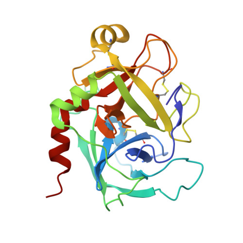
    Hereditary deficiency of factor IXa (fIXa), a key enzyme in blood coagulation, causes hemophilia B, a severe X chromosome-linked bleeding disorder afflicting 1 in 30,000 males; clinical studies have identified nearly 500 deleterious variants. The x-ray structure of porcine fIXa described here shows the atomic origins of the disease, while the spatial distribution of mutation sites suggests a structural model for factor X activation by phospholipid-bound fIXa and cofactor VIIIa. The 3.0-A-resolution diffraction data clearly show the structures of the serine proteinase module and the two preceding epidermal growth factor (EGF)-like modules; the N-terminal Gla module is partially disordered. The catalytic module, with covalent inhibitor D-Phe-1I-Pro-2I-Arg-3I chloromethyl ketone, most closely resembles fXa but differs significantly at several positions. Particularly noteworthy is the strained conformation of Glu-388, a residue strictly conserved in known fIXa sequences but conserved as Gly among other trypsin-like serine proteinases. Flexibility apparent in electron density together with modeling studies suggests that this may cause incomplete active site formation, even after zymogen, and hence the low catalytic activity of fIXa. The principal axes of the oblong EGF-like domains define an angle of 110 degrees, stabilized by a strictly conserved and fIX-specific interdomain salt bridge. The disorder of the Gla module, whose hydrophobic helix is apparent in electron density, can be attributed to the absence of calcium in the crystals; we have modeled the Gla module in its calcium form by using prothrombin fragment 1. The arched module arrangement agrees with fluorescence energy transfer experiments. Most hemophilic mutation sites of surface fIX residues occur on the concave surface of the bent molecule and suggest a plausible model for the membrane-bound ternary fIXa-FVIIIa-fX complex structure: fIXa and an equivalently arranged fX arch across an underlying fVIIIa subdomain from opposite sides; the stabilizing fVIIIa interactions force the catalytic modules together, completing fIXa active site formation and catalytic enhancement.


  • Organizational Affiliation
    • Max-Planck-Institut für Biochemie, Martinsried, Germany.

Macromolecule Content 

  • Total Structure Weight: 43.51 kDa 
  • Atom Count: 3,046 
  • Modeled Residue Count: 381 
  • Deposited Residue Count: 381 
  • Unique protein chains: 2

Macromolecules

Find similar proteins by:|  3D Structure
Entity ID: 1
MoleculeChains  Sequence LengthOrganismDetailsImage
FACTOR IXAA [auth C]235Sus scrofaMutation(s): 0 
Gene Names: PETE
EC: 3.4.21.22
UniProt
Find proteins for P16293 (Sus scrofa)
Explore P16293 
Go to UniProtKB:  P16293
Entity Groups
Sequence Clusters30% Identity50% Identity70% Identity90% Identity95% Identity100% Identity
UniProt GroupP16293
Sequence Annotations
Expand
Reference Sequence
Find similar proteins by:|  3D Structure
Entity ID: 2
MoleculeChains  Sequence LengthOrganismDetailsImage
FACTOR IXAB [auth L]146Sus scrofaMutation(s): 0 
Gene Names: PETE
EC: 3.4.21.22
UniProt
Find proteins for P16293 (Sus scrofa)
Explore P16293 
Go to UniProtKB:  P16293
Entity Groups
Sequence Clusters30% Identity50% Identity70% Identity90% Identity95% Identity100% Identity
UniProt GroupP16293
Sequence Annotations
Expand
Reference Sequence

Small Molecules

Ligands 1 Unique
IDChains Name / Formula / InChI Key2D Diagram3D Interactions
0G6

Query on 0G6


Download:Ideal Coordinates CCD File
C
D-phenylalanyl-N-[(2S,3S)-6-{[amino(iminio)methyl]amino}-1-chloro-2-hydroxyhexan-3-yl]-L-prolinamide
C21 H34 Cl N6 O3
DVFLYEYCMMLBTQ-VSZNYVQBSA-O
Modified Residues  2 Unique
IDChains TypeFormula2D DiagramParent
BHD
Query on BHD
B [auth L]L-PEPTIDE LINKINGC4 H7 N O5ASP
CGU
Query on CGU
B [auth L]L-PEPTIDE LINKINGC6 H9 N O6GLU

Biologically Interesting Molecules (External Reference) 

1 Unique
Entity ID: 3
IDChains NameType/Class2D Diagram3D Interactions
PRD_000020 (0G6)
Query on PRD_000020
C
D-Phe-Pro-Arg-CH2ClPeptide-like / Inhibitor

Experimental Data & Validation

Experimental Data

  • Method: X-RAY DIFFRACTION
  • Resolution: 3.00 Å
  • R-Value Work:  0.198 (Depositor) 
  • R-Value Observed: 0.198 (Depositor) 
Space Group: P 41 21 2
Unit Cell:
Length ( Å )Angle ( ˚ )
a = 128.75α = 90
b = 128.75β = 90
c = 77.2γ = 90
Software Package:
Software NamePurpose
MOSFLMdata reduction
X-PLORmodel building
X-PLORrefinement
X-PLORphasing

Structure Validation

View Full Validation Report


Entry History 

Deposition Data

Revision History  (Full details and data files)

  • Version 1.0: 1996-08-17
    Type: Initial release
  • Version 1.1: 2008-03-24
    Changes: Version format compliance
  • Version 1.2: 2011-07-13
    Changes: Atomic model, Version format compliance
  • Version 1.3: 2012-06-20
    Changes: Non-polymer description
  • Version 1.4: 2013-02-27
    Changes: Other
  • Version 1.5: 2025-03-26
    Changes: Advisory, Data collection, Database references, Derived calculations, Structure summary