1PBO | pdb_00001pbo

COMPLEX OF BOVINE ODORANT BINDING PROTEIN (OBP) WITH A SELENIUM CONTAINING ODORANT


Experimental Data Snapshot

  • Method: X-RAY DIFFRACTION
  • Resolution: 2.20 Å
  • R-Value Work: 
    0.190 (Depositor) 
  • R-Value Observed: 
    0.190 (Depositor) 

wwPDB Validation 3D Report Full Report

Validation slider image for 1PBO

This is version 1.3 of the entry. See complete history

Literature

The three-dimensional structure of bovine odorant binding protein and its mechanism of odor recognition.

Bianchet, M.A.Bains, G.Pelosi, P.Pevsner, J.Snyder, S.H.Monaco, H.L.Amzel, L.M.

(1996) Nat Struct Biol 3: 934-939

  • DOI: https://doi.org/10.1038/nsb1196-934
  • Primary Citation Related Structures: 
    1PBO

  • PubMed Abstract: 

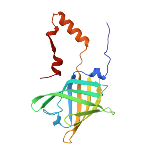
    Odorant binding protein (OBP) is the major odorant binding component of mammalian nasal mucosa. The two structures of bovine OBP reported in this paper (one crystallized as purified and one soaked in the presence of a selenium-containing odorant) show that: (i) the OBP dimer is composed of two compact domains related by an approximate two-fold axis of symmetry; (ii) between residues 122 and 123 the polypeptide chains cross from one domain to the other such that each domain is formed by residues from both monomers; (iii) purified OBP already contains two bound odorant molecules (one per monomer)-odorant binding occurs by replacement of these molecules with the added odorant; and (iv) the structure of the odorant binding site can explain OBP's extraordinarily broad odorant specificity.


  • Organizational Affiliation
    • Department of Biophysics and Biophysical Chemistry, University of Pisa, Italy. mario@neruda.med.jhu.edu

Macromolecule Content 

  • Total Structure Weight: 37.46 kDa 
  • Atom Count: 2,589 
  • Modeled Residue Count: 308 
  • Deposited Residue Count: 318 
  • Unique protein chains: 1

Macromolecules

Find similar proteins by:|  3D Structure
Entity ID: 1
MoleculeChains  Sequence LengthOrganismDetailsImage
ODORANT BINDING PROTEIN
A, B
159Bos taurusMutation(s): 0 
UniProt
Find proteins for P07435 (Bos taurus)
Explore P07435 
Go to UniProtKB:  P07435
Entity Groups
Sequence Clusters30% Identity50% Identity70% Identity90% Identity95% Identity100% Identity
UniProt GroupP07435
Sequence Annotations
Expand
Reference Sequence

Small Molecules

Ligands 1 Unique
IDChains Name / Formula / InChI Key2D Diagram3D Interactions
SES

Query on SES


Download:Ideal Coordinates CCD File
C [auth A],
D [auth B]
4-butyl-5-propyl-1,3-selenazol-2-amine
C10 H18 N2 Se
DOUUMUUUWUAPPC-UHFFFAOYSA-N

Experimental Data & Validation

Experimental Data

  • Method: X-RAY DIFFRACTION
  • Resolution: 2.20 Å
  • R-Value Work:  0.190 (Depositor) 
  • R-Value Observed: 0.190 (Depositor) 
Space Group: P 1 21 1
Unit Cell:
Length ( Å )Angle ( ˚ )
a = 41.87α = 90
b = 65.18β = 98.13
c = 55.54γ = 90
Software Package:
Software NamePurpose
X-PLORmodel building
X-PLORrefinement
XENGENdata reduction
X-PLORphasing

Structure Validation

View Full Validation Report


Entry History 

Deposition Data

Revision History  (Full details and data files)

  • Version 1.0: 1997-07-23
    Type: Initial release
  • Version 1.1: 2008-03-03
    Changes: Version format compliance
  • Version 1.2: 2011-07-13
    Changes: Non-polymer description, Version format compliance
  • Version 1.3: 2024-02-14
    Changes: Data collection, Database references, Derived calculations, Other