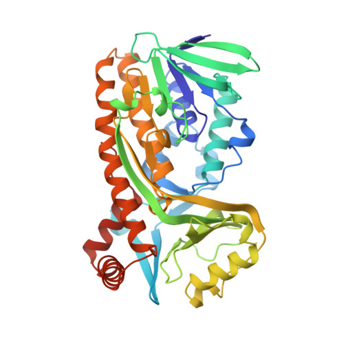
Crystal structures of wild-type p-hydroxybenzoate hydroxylase complexed with 4-aminobenzoate,2,4-dihydroxybenzoate, and 2-hydroxy-4-aminobenzoate and of the Tyr222Ala mutant complexed with 2-hydroxy-4-aminobenzoate. Evidence for a proton channel and a new binding mode of the flavin ring
Schreuder, H.A., Mattevi, A., Obmolova, G., Kalk, K.H., Hol, W.G., van der Bolt, F.J., van Berkel, W.J.(1994) Biochemistry 33: 10161-10170
- PubMed: 7520279 Search on PubMed
- DOI: https://doi.org/10.1021/bi00199a044
- Primary Citation Related Structures:
1PBB, 1PBC, 1PBD, 1PBF - PubMed Abstract:
The crystal structures of wild-type p-hydroxybenzoate hydroxylase from Pseudomonas fluorescens, complexed with the substrate analogues 4-aminobenzoate, 2,4-dihydroxybenzoate, and 2-hydroxy-4-aminobenzoate have been determined at 2.3-, 2.5-, and 2.8-A resolution, respectively. In addition, the crystal structure of a Tyr222Ala mutant, complexed with 2-hydroxy-4-aminobenzoate, has been determined at 2.7-A resolution. The structures have been refined to R factors between 14.5% and 15.8% for data between 8.0 A and the high-resolution limit. The differences between these complexes and the wild-type enzyme-substrate complex are all concentrated in the active site region. Binding of substrate analogues bearing a 4-amino group (4-aminobenzoate and 2-hydroxy-4-aminobenzoate) leads to binding of a water molecule next to the active site Tyr385. As a result, a continuous hydrogen-bonding network is present between the 4-amino group of the substrate analogue and the side chain of His72. It is likely that this hydrogen-bonding network is transiently present during normal catalysis, where it may or may not function as a proton channel assisting the deprotonation of the 4-hydroxyl group of the normal substrate upon binding to the active site. Binding of substrate analogues bearing a hydroxyl group at the 2-position (2,4-dihydroxybenzoate and 2-hydroxy-4-aminobenzoate) leads to displacement of the flavin ring from the active site. The flavin is no longer in the active site (the "in" conformation) but is in the cleft leading to the active site instead (the "out" conformation). It is proposed that movement of the FAD out of the active site may provide an entrance for the substrate to enter the active site and an exit for the product to leave.
- Bioson Research Institute, University of Groningen, The Netherlands.
Organizational Affiliation: