1P9N

Crystal structure of Escherichia coli MobB.


Experimental Data Snapshot

  • Method: X-RAY DIFFRACTION
  • Resolution: 2.80 Å
  • R-Value Free: 
    0.308 (Depositor), 0.323 (DCC) 
  • R-Value Work: 
    0.275 (Depositor), 0.297 (DCC) 
  • R-Value Observed: 
    0.275 (Depositor) 

wwPDB Validation   3D Report Full Report


This is version 1.4 of the entry. See complete history


Literature

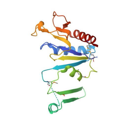
Molecules of Escherichia coli MobB assemble into densely packed hollow cylinders in a crystal lattice with 75% solvent content.

Rangarajan, S.E.Tocilj, A.Li, Y.Iannuzzi, P.Matte, A.Cygler, M.

(2003) Acta Crystallogr D Biol Crystallogr 59: 2348-2352

  • DOI: https://doi.org/10.1107/s090744490301967x
  • Primary Citation of Related Structures:  
    1P9N

  • PubMed Abstract: 

    The crystal structure of Escherichia coli MobB, an enzyme involved in the final step of molybdenum-cofactor biosynthesis, forms intertwined dimers. Each molecule consists of two segments and requires the second monomer for stable folding. Dimerization buries a quarter of the solvent-accessible area of the monomer. These dimers assemble into a hexagonal lattice with P6(4)22 symmetry and occupy only approximately 25% of the unit-cell volume. The symmetry-related dimers associate tightly into a helical structure with a diameter of 250 A and a pitch of 98 A. Two such helices are intertwined, shifted by 49 A along the sixfold axis. Within the crystal, these helices form thin-walled cylinders with an external diameter of 250 A and an internal diameter of 190 A. Their center is filled with solvent. These cylinders pack closely together, forming a hexagonal lattice with the highest possible packing density. This arrangement of dimers allows extensive intermolecular contacts with 75% solvent content in the crystal.


  • Organizational Affiliation

    Department of Biochemistry, McGill University, Canada.


Macromolecules
Find similar proteins by:  (by identity cutoff)  |  3D Structure
Entity ID: 1
MoleculeChains Sequence LengthOrganismDetailsImage
Molybdopterin-guanine dinucleotide biosynthesis protein B
A, B
170Escherichia coliMutation(s): 5 
Gene Names: MobB
UniProt
Find proteins for P32125 (Escherichia coli (strain K12))
Explore P32125 
Go to UniProtKB:  P32125
Entity Groups  
Sequence Clusters30% Identity50% Identity70% Identity90% Identity95% Identity100% Identity
UniProt GroupP32125
Sequence Annotations
Expand
  • Reference Sequence
Small Molecules
Modified Residues  1 Unique
IDChains TypeFormula2D DiagramParent
MSE
Query on MSE
A, B
L-PEPTIDE LINKINGC5 H11 N O2 SeMET
Experimental Data & Validation

Experimental Data

  • Method: X-RAY DIFFRACTION
  • Resolution: 2.80 Å
  • R-Value Free:  0.308 (Depositor), 0.323 (DCC) 
  • R-Value Work:  0.275 (Depositor), 0.297 (DCC) 
  • R-Value Observed: 0.275 (Depositor) 
Space Group: P 64 2 2
Unit Cell:
Length ( Å )Angle ( ˚ )
a = 239.001α = 90
b = 239.001β = 90
c = 49.046γ = 120
Software Package:
Software NamePurpose
CNSrefinement
HKL-2000data reduction
SCALEPACKdata scaling
PHASESphasing

Structure Validation

View Full Validation Report


Entry History 

Revision History  (Full details and data files)

  • Version 1.0: 2003-05-20
    Type: Initial release
  • Version 1.1: 2008-04-29
    Changes: Version format compliance
  • Version 1.2: 2011-07-13
    Changes: Version format compliance
  • Version 1.3: 2018-01-31
    Changes: Experimental preparation
  • Version 1.4: 2024-10-30
    Changes: Data collection, Database references, Derived calculations, Structure summary