1P8Z

Complex Between Rabbit Muscle alpha-Actin: Human Gelsolin Residues Val26-Glu156


Experimental Data Snapshot

  • Method: X-RAY DIFFRACTION
  • Resolution: 2.60 Å
  • R-Value Free: 0.263 
  • R-Value Work: 0.208 
  • R-Value Observed: 0.208 

wwPDB Validation   3D Report Full Report


Ligand Structure Quality Assessment 


This is version 1.2 of the entry. See complete history


Literature

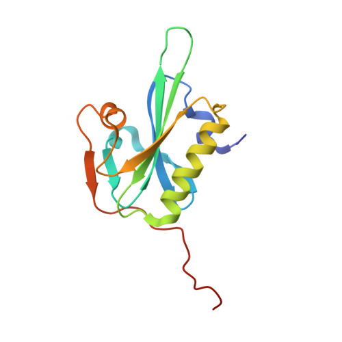
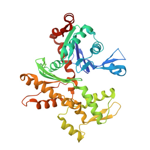
From the First to the second domain of gelsolin: A common path on the surface of actin?

Irobi, E.Burtnick, L.D.Urosev, D.Narayan, K.Robinson, R.C.

(2003) FEBS Lett 552: 86-90

  • DOI: https://doi.org/10.1016/s0014-5793(03)00934-7
  • Primary Citation of Related Structures:  
    1P8Z

  • PubMed Abstract: 
  • We present the 2.6 A resolution crystal structure of a complex formed between G-actin and gelsolin fragment Met25-Gln160 (G1+). The structure differs from those of other gelsolin domain 1 (G1) complexes in that an additional six amino acid residues from the crucial linker region into gelsolin domain 2 (G2) are visible and are attached securely to the surface of actin ...

    We present the 2.6 A resolution crystal structure of a complex formed between G-actin and gelsolin fragment Met25-Gln160 (G1+). The structure differs from those of other gelsolin domain 1 (G1) complexes in that an additional six amino acid residues from the crucial linker region into gelsolin domain 2 (G2) are visible and are attached securely to the surface of actin. The linker segment extends away from G1 up the face of actin in a direction that infers G2 will bind along the same long-pitch helical strand as the actin bound to G1. This is consistent with a mechanism whereby G2 attaches gelsolin to the side of a filament and then directs G1 toward a position where it would disrupt actin-actin contacts. Alignment of the sequence of the structurally important residues within the G1-G2 linker with those of WH2 (WASp homology domain 2) domain protein family members (e.g. WASp (Wiscott-Aldridge syndrome protein) and thymosin beta4) suggests that the opposing activities of filament assembly and disassembly may exploit a common patch on the surface of actin.


    Organizational Affiliation

    Department of Medical Biochemistry and Microbiology, Uppsala University, Box 582, 751 23 Uppsala, Sweden.


Macromolecules
Find similar proteins by:  (by identity cutoff)  |  3D Structure
Entity ID: 1
MoleculeChainsSequence LengthOrganismDetailsImage
Gelsolin precursor, plasmaA [auth G]136Homo sapiensMutation(s): 0 
Gene Names: GSN
UniProt & NIH Common Fund Data Resources
Find proteins for P06396 (Homo sapiens)
Explore P06396 
Go to UniProtKB:  P06396
PHAROS:  P06396
Entity Groups  
Sequence Clusters30% Identity50% Identity70% Identity90% Identity95% Identity100% Identity
UniProt GroupP06396
Protein Feature View
Expand
  • Reference Sequence
Find similar proteins by:  (by identity cutoff)  |  3D Structure
Entity ID: 2
MoleculeChainsSequence LengthOrganismDetailsImage
Actin, alpha skeletal muscleB [auth A]377Oryctolagus cuniculusMutation(s): 0 
Gene Names: ACTA1ACTA
UniProt
Find proteins for P68135 (Oryctolagus cuniculus)
Explore P68135 
Go to UniProtKB:  P68135
Entity Groups  
Sequence Clusters30% Identity50% Identity70% Identity90% Identity95% Identity100% Identity
UniProt GroupP68135
Protein Feature View
Expand
  • Reference Sequence
Small Molecules
Ligands 3 Unique
IDChainsName / Formula / InChI Key2D Diagram3D Interactions
ATP
Query on ATP

Download Ideal Coordinates CCD File 
G [auth A]ADENOSINE-5'-TRIPHOSPHATE
C10 H16 N5 O13 P3
ZKHQWZAMYRWXGA-KQYNXXCUSA-N
 Ligand Interaction
CD
Query on CD

Download Ideal Coordinates CCD File 
C [auth G],
E [auth A],
F [auth A]
CADMIUM ION
Cd
WLZRMCYVCSSEQC-UHFFFAOYSA-N
 Ligand Interaction
CA
Query on CA

Download Ideal Coordinates CCD File 
D [auth G]CALCIUM ION
Ca
BHPQYMZQTOCNFJ-UHFFFAOYSA-N
 Ligand Interaction
Experimental Data & Validation

Experimental Data

  • Method: X-RAY DIFFRACTION
  • Resolution: 2.60 Å
  • R-Value Free: 0.263 
  • R-Value Work: 0.208 
  • R-Value Observed: 0.208 
  • Space Group: P 31 1 2
Unit Cell:
Length ( Å )Angle ( ˚ )
a = 114.22α = 90
b = 114.22β = 90
c = 93.77γ = 120
Software Package:
Software NamePurpose
DENZOdata reduction
SCALEPACKdata scaling
AMoREphasing
CNSrefinement

Structure Validation

View Full Validation Report


Ligand Structure Quality Assessment 


Entry History 

Deposition Data

Revision History  (Full details and data files)

  • Version 1.0: 2003-10-14
    Type: Initial release
  • Version 1.1: 2008-04-29
    Changes: Version format compliance
  • Version 1.2: 2011-07-13
    Changes: Version format compliance