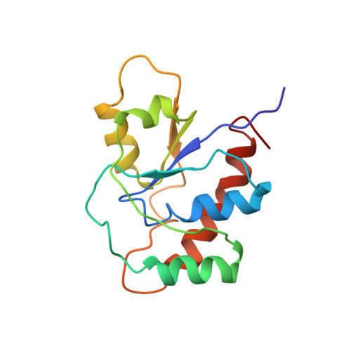
Solution structure of the low-molecular-weight protein tyrosine phosphatase from Tritrichomonas foetus reveals a flexible phosphate binding loop.
Gustafson, C.L., Stauffacher, C.V., Hallenga, K., Van Etten, R.L.(2005) Protein Sci 14: 2515-2525
- PubMed: 16195543 Search on PubMedSearch on PubMed Central
- DOI: https://doi.org/10.1110/ps.051618805
- Primary Citation Related Structures:
1P8A - PubMed Abstract:
Eukaryotic low-molecular-weight protein tyrosine phosphatases (LMW PTPs) contain a conserved serine, a histidine with an elevated pKa, and an active site asparagine that together form a highly conserved hydrogen bonding network. This network stabilizes the active site phosphate binding loop for optimal substrate binding and catalysis. In the phosphatase from the bovine parasite Tritrichomonas foetus (TPTP), both the conserved serine (S37) and asparagine (N14) are present, but the conserved histidine has been replaced by a glutamine residue (Q67). Site-directed mutagenesis, kinetic, and spectroscopic experiments suggest that Q67 is located near the active site and is important for optimal catalytic activity. Kinetic experiments also suggest that S37 participates in the active site/hydrogen bonding network. Nuclear magnetic resonance spectroscopy was used to determine the three-dimensional structure of the TPTP enzyme and to further examine the roles of S37 and Q67. The backbone conformation of the TPTP phosphate binding loop is nearly superimposable with that of other tyrosine phosphatases, with N14 existing in a strained, left-handed conformation that is a hallmark of the active site hydrogen bonding network in the LMW PTPs. As expected, both S37 and Q67 are located at the active site, but in the consensus structure they are not within hydrogen bonding distance of N14. The hydrogen bond interactions that are observed in X-ray structures of LMW PTPs may in fact be transient in solution. Protein dynamics within the active site hydrogen bonding network appear to be affected by the presence of substrate or bound inhibitors such as inorganic phosphate.
- Department of Chemistry, Purdue University, West Lafayette, IN 47907-2084, USA.
Organizational Affiliation: