1OYB | pdb_00001oyb

OLD YELLOW ENZYME AT 2 ANGSTROMS RESOLUTION: OVERALL STRUCTURE, LIGAND BINDING AND COMPARISON WITH RELATED FLAVOPROTEINS


Experimental Data Snapshot

  • Method: X-RAY DIFFRACTION
  • Resolution: 2.00 Å
  • R-Value Free: 
    0.236 (Depositor) 
  • R-Value Work: 
    0.173 (Depositor) 
  • R-Value Observed: 
    0.173 (Depositor) 

wwPDB Validation 3D Report Full Report

Validation slider image for 1OYB

This is version 1.3 of the entry. See complete history

Literature

Old yellow enzyme at 2 A resolution: overall structure, ligand binding, and comparison with related flavoproteins.

Fox, K.M.Karplus, P.A.

(1994) Structure 2: 1089-1105

  • Primary Citation Related Structures: 
    1OYA, 1OYB, 1OYC

  • PubMed Abstract: 

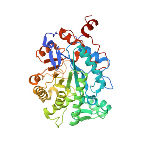
    Old yellow enzyme (OYE) was the first flavoenzyme purified, but its function is still unknown. Nevertheless, the NADPH oxidase activity, the flavin mononucleotide environment and the ligand-binding properties of OYE have been extensively studied by biochemical and spectroscopic approaches. Full interpretation of these data requires structural information. The crystal structures of oxidized and reduced OYE at 2 A resolution reveal an alpha/beta-barrel topology clearly related to trimethylamine dehydrogenase. Complexes of OYE with p-hydroxybenzaldehyde, beta-estradiol, and an NADPH analog show all three binding at a common site, stacked on the flavin. The putative NADPH binding mode is novel as it involves primary recognition of the nicotinamide mononucleotide portion. This work shows that the striking spectral changes seen upon phenol binding are due to close physical association of the flavin and phenolate. It also identifies the structural class of OYE and suggests that if NADPH is its true substrate, then OYE has adopted NADPH dependence during evolution.


  • Organizational Affiliation
    • Section of Biochemistry, Molecular and Cell Biology, Cornell University, Ithaca, New York 14853.

Macromolecule Content 

  • Total Structure Weight: 45.65 kDa 
  • Atom Count: 3,344 
  • Modeled Residue Count: 399 
  • Deposited Residue Count: 400 
  • Unique protein chains: 1

Macromolecules

Find similar proteins by:|  3D Structure
Entity ID: 1
MoleculeChains  Sequence LengthOrganismDetailsImage
OLD YELLOW ENZYME400Saccharomyces pastorianusMutation(s): 0 
Gene Names: OYE1
EC: 1.6.99.1
UniProt
Find proteins for Q02899 (Saccharomyces pastorianus)
Explore Q02899 
Go to UniProtKB:  Q02899
Entity Groups
Sequence Clusters30% Identity50% Identity70% Identity90% Identity95% Identity100% Identity
UniProt GroupQ02899
Sequence Annotations
Expand
Reference Sequence

Small Molecules

Ligands 2 Unique
IDChains Name / Formula / InChI Key2D Diagram3D Interactions
FMN

Query on FMN


Download:Ideal Coordinates CCD File
B [auth A]FLAVIN MONONUCLEOTIDE
C17 H21 N4 O9 P
FVTCRASFADXXNN-SCRDCRAPSA-N
HBA

Query on HBA


Download:Ideal Coordinates CCD File
C [auth A]P-HYDROXYBENZALDEHYDE
C7 H6 O2
RGHHSNMVTDWUBI-UHFFFAOYSA-N

Experimental Data & Validation

Experimental Data

  • Method: X-RAY DIFFRACTION
  • Resolution: 2.00 Å
  • R-Value Free:  0.236 (Depositor) 
  • R-Value Work:  0.173 (Depositor) 
  • R-Value Observed: 0.173 (Depositor) 
Space Group: P 43 21 2
Unit Cell:
Length ( Å )Angle ( ˚ )
a = 142.88α = 90
b = 142.88β = 90
c = 43.01γ = 90
Software Package:
Software NamePurpose
X-PLORmodel building
X-PLORrefinement
X-PLORphasing

Structure Validation

View Full Validation Report


Entry History 

Deposition Data

Revision History  (Full details and data files)

  • Version 1.0: 1994-11-30
    Type: Initial release
  • Version 1.1: 2008-03-24
    Changes: Version format compliance
  • Version 1.2: 2011-07-13
    Changes: Version format compliance
  • Version 1.3: 2024-02-14
    Changes: Data collection, Database references, Derived calculations, Other