Crystallographic analysis of oxygenated and deoxygenated states of arthropod hemocyanin shows unusual differences.
Magnus, K.A., Hazes, B., Ton-That, H., Bonaventura, C., Bonaventura, J., Hol, W.G.(1994) Proteins 19: 302-309
- PubMed: 7984626 Search on PubMed
- DOI: https://doi.org/10.1002/prot.340190405
- Primary Citation Related Structures:
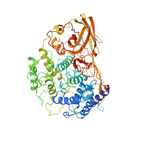
1OXY - PubMed Abstract:
The X-ray structure of an oxygenated hemocyanin molecule, subunit II of Limulus polyphemus hemocyanin, was determined at 2.4 A resolution and refined to a crystallographic R-factor of 17.1%. The 73-kDa subunit crystallizes with the symmetry of the space group R32 with one subunit per asymmetric unit forming hexamers with 32 point group symmetry. Molecular oxygen is bound to a dinuclear copper center in the protein's second domain, symmetrically between and equidistant from the two copper atoms. The copper-copper distance in oxygenated Limulus hemocyanin is 3.6 +/- 0.2 A, which is surprisingly 1 A less than that seen previously in deoxygenated Limulus polyphemus subunit II hemocyanin (Hazes et al., Protein Sci. 2:597, 1993). Away from the oxygen binding sites, the tertiary and quaternary structures of oxygenated and deoxygenated Limulus subunit II hemocyanins are quite similar. A major difference in tertiary structures is seen, however, when the Limulus structures are compared with deoxygenated Panulirus interruptus hemocyanin (Volbeda, A., Hol, W.G.J.J. Mol. Biol. 209:249, 1989) where the position of domain 1 is rotated by 8 degrees with respect to domains 2 and 3. We postulate this rotation plays an important role in cooperativity and regulation of oxygen affinity in all arthropod hemocyanins.
- Department of Biochemistry, Case Western Reserve University, School of Medicine, Cleveland, Ohio 44106-4935.
Organizational Affiliation: