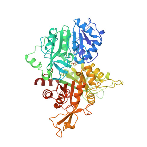
1OX5

TOWARDS UNDERSTANDING THE MECHANISM OF THE COMPLEX CYCLIZATION REACTION CATALYZED BY IMIDAZOLE GLYCEROPHOSPHATE SYNTHASE


Experimental Data Snapshot

  • Method: X-RAY DIFFRACTION
  • Resolution: 2.50 Å
  • R-Value Free: 0.246 
  • R-Value Work: 0.223 

Starting Model: experimental
View more details

wwPDB Validation   3D Report Full Report


Ligand Structure Quality Assessment 


This is version 1.3 of the entry. See complete history


Literature

Towards Understanding the Mechanism of the Complex Cyclization Reaction Catalyzed by Imidazole Glycerophosphate Synthase:Crystal Structures of a Ternary Complex and the Free Enzyme

Chaudhuri, B.N.Lange, S.C.Myers, R.S.Davisson, V.J.Smith, J.L.

(2003) Biochemistry 42: 7003-7012

  • DOI: https://doi.org/10.1021/bi034320h
  • Primary Citation of Related Structures:  
    1OX4, 1OX5, 1OX6

  • PubMed Abstract: 

    Imidazole glycerol phosphate synthase catalyzes formation of the imidazole ring in histidine biosynthesis. The enzyme is also a glutamine amidotransferase, which produces ammonia in a glutaminase active site and channels it through a 30-A internal tunnel to a cyclase active site. Glutaminase activity is impaired in the resting enzyme, and stimulated by substrate binding in the cyclase active site. The signaling mechanism was investigated in the crystal structure of a ternary complex in which the glutaminase active site was inactivated by a glutamine analogue and the unstable cyclase substrate was cryo-trapped in the active site. The orientation of N(1)-(5'-phosphoribulosyl)-formimino-5-aminoimidazole-4-carboxamide ribonucleotide in the cyclase active site implicates one side of the cyclase domain in signaling to the glutaminase domain. This side of the cyclase domain contains the interdomain hinge. Two interdomain hydrogen bonds, which do not exist in more open forms of the enzyme, are proposed as molecular signals. One hydrogen bond connects the cyclase domain to the substrate analogue in the glutaminase active site. The second hydrogen bond connects to a peptide that forms an oxyanion hole for stabilization of transient negative charge during glutamine hydrolysis. Peptide rearrangement induced by a fully closed domain interface is proposed to activate the glutaminase by unblocking the oxyanion hole. This interpretation is consistent with biochemical results [Myers, R. S., et al., (2003) Biochemistry 42, 7013-7022, the accompanying paper in this issue] and with structures of the free enzyme and a binary complex with a second glutamine analogue.


  • Organizational Affiliation

    Department of Biological Sciences, Purdue University, West Lafayette, Indiana 47907, USA.


Macromolecules
Find similar proteins by:  (by identity cutoff)  |  3D Structure
Entity ID: 1
MoleculeChains Sequence LengthOrganismDetailsImage
Imidazole glycerol phosphate synthase hisHF
A, B
555Saccharomyces cerevisiaeMutation(s): 1 
Gene Names: HIS7
EC: 2.4.2 (PDB Primary Data), 4.1.3 (PDB Primary Data), 4.3.2.10 (UniProt), 3.5.1.2 (UniProt)
UniProt
Find proteins for P33734 (Saccharomyces cerevisiae (strain ATCC 204508 / S288c))
Explore P33734 
Go to UniProtKB:  P33734
Entity Groups  
Sequence Clusters30% Identity50% Identity70% Identity90% Identity95% Identity100% Identity
UniProt GroupP33734
Sequence Annotations
Expand
  • Reference Sequence
Small Molecules
Ligands 2 Unique
IDChains Name / Formula / InChI Key2D Diagram3D Interactions
1PR
Query on 1PR

Download Ideal Coordinates CCD File 
D [auth A],
E [auth B]
PHOSPHORIC ACID MONO-[5-({[5-CARBAMOYL-3-(5-PHOSPHONOOXY-5-DEOXY-RIBOFURANOSYL)- 3H-IMIDAZOL-4-YLAMINO]-METHYL}-AMINO)-2,3,4-TRIHYDROXY-PENTYL] ESTER
C15 H29 N5 O15 P2
HDCXLRQQJHBDSI-DYHIIFNWSA-N
NI
Query on NI

Download Ideal Coordinates CCD File 
C [auth A]NICKEL (II) ION
Ni
VEQPNABPJHWNSG-UHFFFAOYSA-N
Modified Residues  1 Unique
IDChains TypeFormula2D DiagramParent
5CS
Query on 5CS
A, B
L-PEPTIDE LINKINGC8 H15 N3 O5 SCYS
Experimental Data & Validation

Experimental Data

  • Method: X-RAY DIFFRACTION
  • Resolution: 2.50 Å
  • R-Value Free: 0.246 
  • R-Value Work: 0.223 
  • Space Group: P 21 21 21
Unit Cell:
Length ( Å )Angle ( ˚ )
a = 98.8α = 90
b = 111.5β = 90
c = 117.2γ = 90
Software Package:
Software NamePurpose
HKL-2000data scaling
CNSrefinement

Structure Validation

View Full Validation Report


Ligand Structure Quality Assessment 


Entry History 

Deposition Data

Revision History  (Full details and data files)

  • Version 1.0: 2003-06-17
    Type: Initial release
  • Version 1.1: 2008-04-29
    Changes: Version format compliance
  • Version 1.2: 2011-07-13
    Changes: Version format compliance
  • Version 1.3: 2023-08-16
    Changes: Data collection, Database references, Derived calculations, Refinement description