A conserved structural motif reveals the essential transcriptional repression function of Spen proteins and their role in developmental signaling
Schwabe, J.W., Ariyoshi, M.(2003) Genes Dev 17: 1909-1920
- PubMed: 12897056 Search on PubMedSearch on PubMed Central
- DOI: https://doi.org/10.1101/gad.266203
- Primary Citation Related Structures:
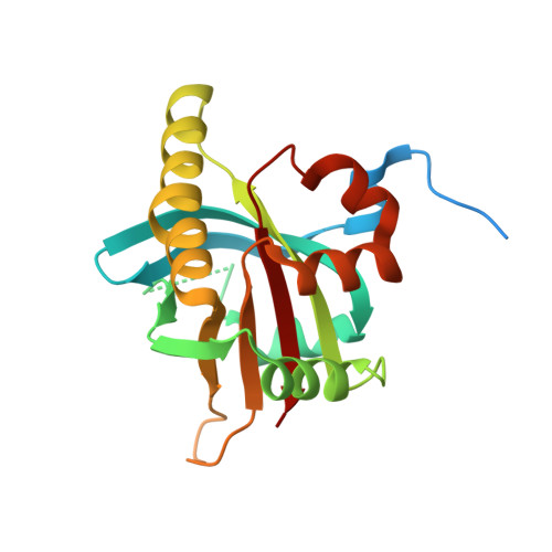
1OW1 - PubMed Abstract:
Spen proteins regulate the expression of key transcriptional effectors in diverse signaling pathways. They are large proteins characterized by N-terminal RNA-binding motifs and a highly conserved C-terminal SPOC domain. The specific biological role of the SPOC domain (Spen paralog and ortholog C-terminal domain), and hence, the common function of Spen proteins, has been unclear to date. The Spen protein, SHARP (SMRT/HDAC1-associated repressor protein), was identified as a component of transcriptional repression complexes in both nuclear receptor and Notch/RBP-Jkappa signaling pathways. We have determined the 1.8 A crystal structure of the SPOC domain from SHARP. This structure shows that essentially all of the conserved surface residues map to a positively charged patch. Structure-based mutational analysis indicates that this conserved region is responsible for the interaction between SHARP and the universal transcriptional corepressor SMRT/NCoR (silencing mediator for retinoid and thyroid receptors/nuclear receptor corepressor. We demonstrate that this interaction involves a highly conserved acidic motif at the C terminus of SMRT/NCoR. These findings suggest that the conserved function of the SPOC domain is to mediate interaction with SMRT/NCoR corepressors, and that Spen proteins play an essential role in the repression complex.
- Medical Research Council, Laboratory of Molecular Biology, Cambridge CB2 2QH, UK.
Organizational Affiliation: