1OOW

The crystal structure of the spinach plastocyanin double mutant G8D/L12E gives insight into its low reactivity towards photosystem 1 and cytochrome f


Experimental Data Snapshot

  • Method: X-RAY DIFFRACTION
  • Resolution: 2.00 Å
  • R-Value Free: 0.232 
  • R-Value Work: 0.183 
  • R-Value Observed: 0.188 

wwPDB Validation   3D Report Full Report


This is version 1.6 of the entry. See complete history


Literature

The crystal structure of the spinach plastocyanin double mutant G8D/L12E gives insight into its low reactivity towards photosystem 1 and cytochrome f.

Jansson, H.Okvist, M.Jacobson, F.Ejdeback, M.Hansson, O.Sjolin, L.

(2003) Biochim Biophys Acta 1607: 203-210

  • DOI: https://doi.org/10.1016/j.bbabio.2003.09.011
  • Primary Citation of Related Structures:  
    1OOW

  • PubMed Abstract: 

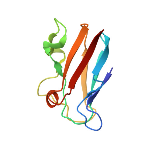
    Plastocyanin (Pc) is a copper-containing protein, which functions as an electron carrier between the cytochrome b(6)f and photosystem 1 (PS1) complexes in the photosynthetic electron transfer (ET) chain. The ET is mediated by His87 situated in the hydrophobic surface in the north region of Pc. Also situated in this region is Leu12, which mutated to other amino acids severely disturbs the ET from cytochrome f and to PS1, indicating the importance of the hydrophobic surface. The crystal structure of the Pc double mutant G8D/L12E has been determined to 2.0 A resolution, with a crystallographic R-factor of 18.3% (R(free)=23.2%). A comparison with the wild-type structure reveals that structural differences are limited to the sites of the mutations. In particular, there is a small but significant change in the hydrophobic surface close to His87. Evidently, this leads to a mismatch in the reactive complex with the redox partners. For PS1 this results in a 20 times weaker binding and an eightfold slower ET as determined by kinetic measurements. The mutations that have been introduced do not affect the optical absorption spectrum. However, there is a small change in the EPR spectrum, which can be related to changes in the copper coordination geometry.


  • Organizational Affiliation

    Center for Structural Biology and Department of Biochemistry and Biophysics, Göteborg University, Box 462, SE-405 30, Göteborg, Sweden.


Macromolecules
Find similar proteins by:  (by identity cutoff)  |  3D Structure
Entity ID: 1
MoleculeChains Sequence LengthOrganismDetailsImage
Plastocyanin, chloroplast99Spinacia oleraceaMutation(s): 2 
Gene Names: PETE
UniProt
Find proteins for P00289 (Spinacia oleracea)
Explore P00289 
Go to UniProtKB:  P00289
Entity Groups  
Sequence Clusters30% Identity50% Identity70% Identity90% Identity95% Identity100% Identity
UniProt GroupP00289
Sequence Annotations
Expand
  • Reference Sequence
Small Molecules
Ligands 1 Unique
IDChains Name / Formula / InChI Key2D Diagram3D Interactions
CU
Query on CU

Download Ideal Coordinates CCD File 
B [auth A]COPPER (II) ION
Cu
JPVYNHNXODAKFH-UHFFFAOYSA-N
Experimental Data & Validation

Experimental Data

  • Method: X-RAY DIFFRACTION
  • Resolution: 2.00 Å
  • R-Value Free: 0.232 
  • R-Value Work: 0.183 
  • R-Value Observed: 0.188 
  • Space Group: P 31 2 1
Unit Cell:
Length ( Å )Angle ( ˚ )
a = 74.761α = 90
b = 74.761β = 90
c = 31.383γ = 120
Software Package:
Software NamePurpose
REFMACrefinement
XDSdata reduction
XSCALEdata scaling

Structure Validation

View Full Validation Report


Entry History 

Deposition Data

Revision History  (Full details and data files)

  • Version 1.0: 2004-02-17
    Type: Initial release
  • Version 1.1: 2008-04-29
    Changes: Version format compliance
  • Version 1.2: 2011-07-13
    Changes: Version format compliance
  • Version 1.3: 2018-03-07
    Changes: Data collection
  • Version 1.4: 2018-10-03
    Changes: Data collection, Derived calculations
  • Version 1.5: 2021-10-27
    Changes: Database references, Derived calculations
  • Version 1.6: 2023-08-16
    Changes: Data collection, Refinement description