Structure of oncomodulin refined at 1.85 A resolution. An example of extensive molecular aggregation via Ca2+.
Ahmed, F.R., Przybylska, M., Rose, D.R., Birnbaum, G.I., Pippy, M.E., MacManus, J.P.(1990) J Mol Biology 216: 127-140
- PubMed: 2231727 Search on PubMed
- DOI: https://doi.org/10.1016/S0022-2836(05)80065-8
- Primary Citation Related Structures:
1OMD - PubMed Abstract:
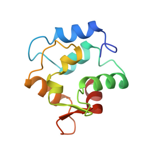
The crystal structure of oncomodulin, a 12,000 Mr protein isolated from rat tumours, has been determined by molecular replacement using the carp parvalbumin structure as a starting model. Refinement was performed by cycles of molecular fitting and restrained least-squares, using area-detector intensity data to 1.85 A resolution. For the 5770 reflections in the range 6.0 to 1.85 A, which were used in the refinement, the crystallographic R-factor is 0.166. The refined model includes residues 2 to 108, three Ca2+ and 87 water molecules per oncomodulin molecule. The oncomodulin backbone is closely related to that of parvalbumin; however, some differences are found after a least-squares fit of the two backbones, with root-mean-square (r.m.s.) deviations of 1 to 2 A in residues 2 to 6, 59 to 61 of the CD loop, 87, 90 and 108. The overall r.m.s. deviation of the backbone residues 5 to 108 is 0.62 A. Each of the two Ca2+ atoms that are bound to the CD and EF loops is co-ordinated to seven oxygen atoms, including one water molecule. The third Ca2+ is also seven-co-ordinated, to five oxygen atoms belonging to three different oncomodulin molecules and to two water molecules which form hydrogen bonds to a fourth oncomodulin; thus, this intermolecular Ca2+ and its equivalents interlink the molecules into zigzag layers normal to the b axis with a spacing of b/2 or 32.14 A. No such extensive molecular aggregation has been reported for any of the related Ca-binding regulatory proteins of the troponin-C family studied thus far. The Ca-O distances in all three polyhedra are in the range 2.07 A to 2.64 A, indicating tightly bound Ca polyhedra.
- Division of Biological Sciences, National Research Council of Canada, Ottawa.
Organizational Affiliation: