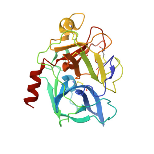
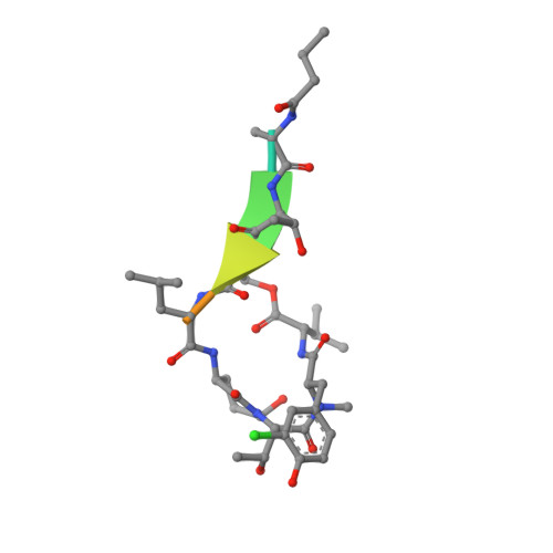
1OKX

Binding Structure of Elastase Inhibitor Scyptolin A


Experimental Data Snapshot

  • Method: X-RAY DIFFRACTION
  • Resolution: 2.80 Å
  • R-Value Free: 0.260 
  • R-Value Work: 0.210 
  • R-Value Observed: 0.210 

Starting Model: experimental
View more details

wwPDB Validation   3D Report Full Report


This is version 1.5 of the entry. See complete history


Literature

Binding Structure of Elastase Inhibitor Scyptolin A

Matern, U.Schleberger, C.Jelakovic, S.Weckesser, J.Schulz, G.E.

(2003) Chem Biol 10: 997

  • DOI: https://doi.org/10.1016/j.chembiol.2003.10.001
  • Primary Citation of Related Structures:  
    1OKX

  • PubMed Abstract: 

    Natural bioactive compounds are of general interest to pharmaceutical research because they may be used as leads in drug development campaigns. Among them, scyptolin A and B from Scytonema hofmanni PCC 7110 are known to inhibit porcine pancreatic elastase, which in turn resembles the attractive drug target neutrophil elastase. The crystal structure of scyptolin A as bound to pancreatic elastase was solved at 2.8 A resolution. The inhibitor occupies the most prominent subsites S1 through S4 of the elastase and prevents a hydrolytic attack by covering the active center with its rigid ring structure. The observed binding structure may help to design potent elastase inhibitors.


  • Organizational Affiliation

    Institut für Biologie II, Mikrobiologie, Albert-Ludwigs-Universität, Schänzlestrasse 1, D-79104 Freiburg im Breisgau, Germany.


Macromolecules
Find similar proteins by:  (by identity cutoff)  |  3D Structure
Entity ID: 1
MoleculeChains Sequence LengthOrganismDetailsImage
ELASTASE 1
A, B
240Sus scrofaMutation(s): 0 
EC: 3.4.21.36
UniProt
Find proteins for P00772 (Sus scrofa)
Explore P00772 
Go to UniProtKB:  P00772
Entity Groups  
Sequence Clusters30% Identity50% Identity70% Identity90% Identity95% Identity100% Identity
UniProt GroupP00772
Sequence Annotations
Expand
  • Reference Sequence

Find similar proteins by:  Sequence   |   3D Structure  

Entity ID: 2
MoleculeChains Sequence LengthOrganismDetailsImage
SCYPTOLIN A
C, D
8Scytonema hofmanniiMutation(s): 0 
Sequence Annotations
Expand
  • Reference Sequence
Biologically Interesting Molecules (External Reference) 1 Unique
Experimental Data & Validation

Experimental Data

  • Method: X-RAY DIFFRACTION
  • Resolution: 2.80 Å
  • R-Value Free: 0.260 
  • R-Value Work: 0.210 
  • R-Value Observed: 0.210 
  • Space Group: P 6 2 2
Unit Cell:
Length ( Å )Angle ( ˚ )
a = 155.89α = 90
b = 155.89β = 90
c = 91.03γ = 120
Software Package:
Software NamePurpose
CNSrefinement
XDSdata reduction
XSCALEdata scaling
MOLREPphasing

Structure Validation

View Full Validation Report


Entry History 

Deposition Data

Revision History  (Full details and data files)

  • Version 1.0: 2003-10-24
    Type: Initial release
  • Version 1.1: 2011-07-13
    Changes: Atomic model, Database references, Derived calculations, Non-polymer description, Structure summary, Version format compliance
  • Version 1.2: 2012-11-30
    Changes: Other
  • Version 1.3: 2018-06-13
    Changes: Data collection, Structure summary
  • Version 1.4: 2019-07-31
    Changes: Advisory, Data collection, Derived calculations
  • Version 1.5: 2023-12-13
    Changes: Data collection, Database references, Derived calculations, Other, Refinement description