1OFX

CRYSTAL STRUCTURE OF AN OKAZAKI FRAGMENT AT 2 ANGSTROMS RESOLUTION


Experimental Data Snapshot

  • Method: X-RAY DIFFRACTION
  • Resolution: 2.00 Å
  • R-Value Observed: 0.156 

wwPDB Validation   3D Report Full Report


This is version 1.3 of the entry. See complete history


Literature

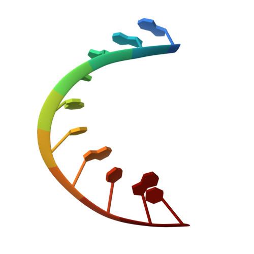
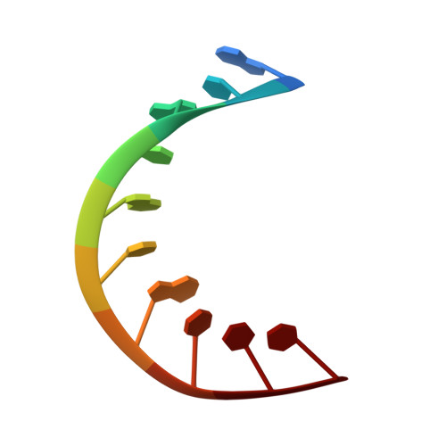
Crystal structure of an Okazaki fragment at 2-A resolution.

Egli, M.Usman, N.Zhang, S.G.Rich, A.

(1992) Proc Natl Acad Sci U S A 89: 534-538

  • DOI: https://doi.org/10.1073/pnas.89.2.534
  • Primary Citation of Related Structures:  
    1OFX

  • PubMed Abstract: 

    In DNA replication, Okazaki fragments are formed as double-stranded intermediates during synthesis of the lagging strand. They are composed of the growing DNA strand primed by RNA and the template strand. The DNA oligonucleotide d(GGGTATACGC) and the chimeric RNA-DNA oligonucleotide r(GCG)d(TATACCC) were combined to form a synthetic Okazaki fragment and its three-dimensional structure was determined by x-ray crystallography. The fragment adopts an overall A-type conformation with 11 residues per turn. Although the base-pair geometry, particularly in the central TATA part, is distorted, there is no evidence for a transition from the A- to the B-type conformation at the junction between RNA.DNA hybrid and DNA duplex. The RNA trimer may, therefore, lock the complete fragment in an A-type conformation.


  • Organizational Affiliation

    Department of Biology, Massachusetts Institute of Technology, Cambridge 02139.


Macromolecules

Find similar nucleic acids by:  Sequence   |   3D Structure  

Entity ID: 1
MoleculeChains LengthOrganismImage
DNA (5'-D(*GP*GP*GP*TP*AP*TP*AP*CP*GP*C)-3')10N/A
Sequence Annotations
Expand
  • Reference Sequence

Find similar nucleic acids by:  Sequence   |   3D Structure  

Entity ID: 2
MoleculeChains LengthOrganismImage
DNA/RNA (5'-R(*GP*CP*GP*)-D(*TP*AP*TP*AP*CP*CP*C)-3')10N/A
Sequence Annotations
Expand
  • Reference Sequence
Small Molecules
Ligands 1 Unique
IDChains Name / Formula / InChI Key2D Diagram3D Interactions
SPM
Query on SPM

Download Ideal Coordinates CCD File 
C [auth B]SPERMINE
C10 H26 N4
PFNFFQXMRSDOHW-UHFFFAOYSA-N
Experimental Data & Validation

Experimental Data

  • Method: X-RAY DIFFRACTION
  • Resolution: 2.00 Å
  • R-Value Observed: 0.156 
  • Space Group: P 21 21 21
Unit Cell:
Length ( Å )Angle ( ˚ )
a = 24.03α = 90
b = 43.67β = 90
c = 48.95γ = 90
Software Package:
Software NamePurpose
PROLSQrefinement
MODIFIEDrefinement

Structure Validation

View Full Validation Report


Entry History 

Deposition Data

Revision History  (Full details and data files)

  • Version 1.0: 1993-04-15
    Type: Initial release
  • Version 1.1: 2008-05-22
    Changes: Version format compliance
  • Version 1.2: 2011-07-13
    Changes: Version format compliance
  • Version 1.3: 2024-02-14
    Changes: Data collection, Database references, Derived calculations