Pb1 Domain-Mediated Heterodimerization in Nadph Oxidase and Signaling Complexes of Atypical Protein Kinase C with Par6 and P62
Wilson, M.I., Gill, D.J., Perisic, O., Quinn, M.T., Williams, R.L.(2003) Mol Cell 12: 39
- PubMed: 12887891 Search on PubMed
- DOI: https://doi.org/10.1016/s1097-2765(03)00246-6
- Primary Citation Related Structures:
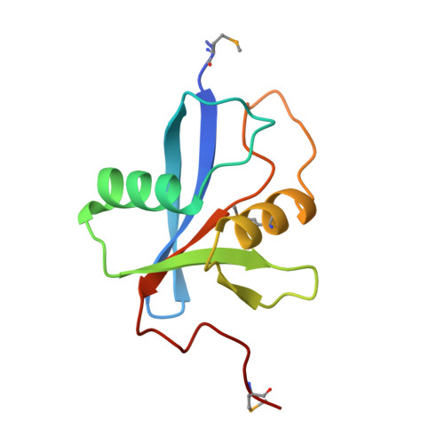
1OEY - PubMed Abstract:
Maximal activation of NADPH oxidase requires formation of a complex between the p40(phox) and p67(phox) subunits via association of their PB1 domains. We have determined the crystal structure of the p40(phox)/p67(phox) PB1 heterodimer, which reveals that both domains have a beta grasp topology and that they bind in a front-to-back arrangement through conserved electrostatic interactions between an acidic OPCA motif on p40(phox) and basic residues in p67(phox). The structure enabled us to identify residues critical for heterodimerization among other members of the PB1 domain family, including the atypical protein kinase C zeta (PKC zeta) and its partners Par6 and p62 (ZIP, sequestosome). Both Par6 and p62 use their basic "back" to interact with the OPCA motif on the "front" of the PKC zeta. Besides heterodimeric interactions, some PB1 domains, like the p62 PB1, can make homotypic front-to-back arrays.
- MRC Laboratory of Molecular Biology, Hills Road, Cambridge CB2 2QH, United Kingdom.
Organizational Affiliation: