1OD8 | pdb_00001od8

Xylanase Xyn10A from Streptomyces lividans in complex with xylobio-isofagomine lactam


Experimental Data Snapshot

  • Method: X-RAY DIFFRACTION
  • Resolution: 1.05 Å
  • R-Value Free: 
    0.126 (Depositor), 0.136 (DCC) 
  • R-Value Work: 
    0.103 (Depositor), 0.119 (DCC) 
  • R-Value Observed: 
    0.104 (Depositor) 

Starting Model: experimental
View more details

wwPDB Validation 3D Report Full Report

Validation slider image for 1OD8

Ligand Structure Quality Assessment 


This is version 2.2 of the entry. See complete history. 

Literature

A Xylobiose-Derived Isofagomine Lactam Glycosidase Inhibitor Binds as its Amide Tautomer

Gloster, T.M., Williams, S.J., Tarling, C., Roberts, S., Dupont, C., Jodoin, P., Shareck, F., Withers, S.G., Davies, G.J.

(2003) Chem Commun (Camb) 8: 944

  • DOI: https://doi.org/10.1039/b301829f
  • Primary Citation Related Structures: 
    1OD8

  • PubMed Abstract: 

    The atomic-resolution structure of a xylobiose-derived isofagomine lactam in complex with the xylanase Xyn10A from Streptomyces lividans reveals that the lactam is bound to the enzyme as the amide tautomer, with "reversed" protonation-states for nucleophile and acid-base.


  • Organizational Affiliation: 
    • Structural Biology Laboratory, Department of Chemistry, The University of York, Heslington, UK.

Macromolecule Content 

  • Total Structure Weight: 34.89 kDa 
  • Atom Count: 3,076 
  • Modeled Residue Count: 302 
  • Deposited Residue Count: 313 
  • Unique protein chains: 1

Macromolecules

Find similar proteins by:|  3D Structure
Entity ID: 1
MoleculeChains  Sequence LengthOrganismDetailsImage
ENDO-1,4-BETA-XYLANASE A313Streptomyces lividansMutation(s): 0 
EC: 3.2.1.8
UniProt
Find proteins for P26514 (Streptomyces lividans)
Explore P26514 
Go to UniProtKB:  P26514
Entity Groups
Sequence Clusters30% Identity50% Identity70% Identity90% Identity95% Identity100% Identity
UniProt GroupP26514
Sequence Annotations
Expand
Reference Sequence

Small Molecules

Ligands 4 Unique
IDChains Name / Formula / InChI Key2D Diagram3D Interactions
XYP

Query on XYP


Download:Ideal Coordinates CCD File
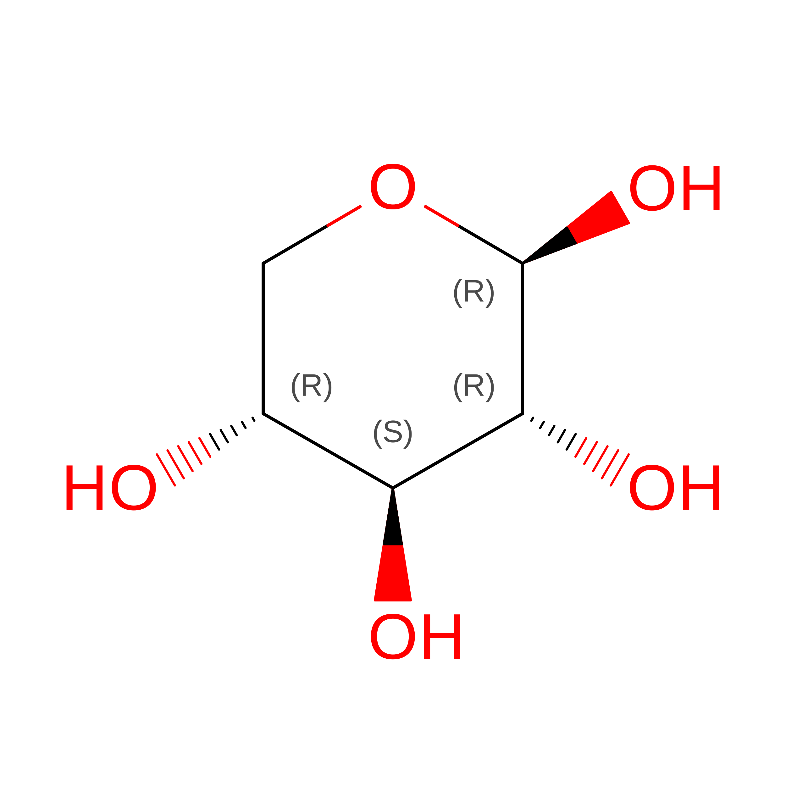
F [auth A],
J [auth A]
beta-D-xylopyranose
C5 H10 O5
SRBFZHDQGSBBOR-KKQCNMDGSA-N
XDL

Query on XDL


Download:Ideal Coordinates CCD File
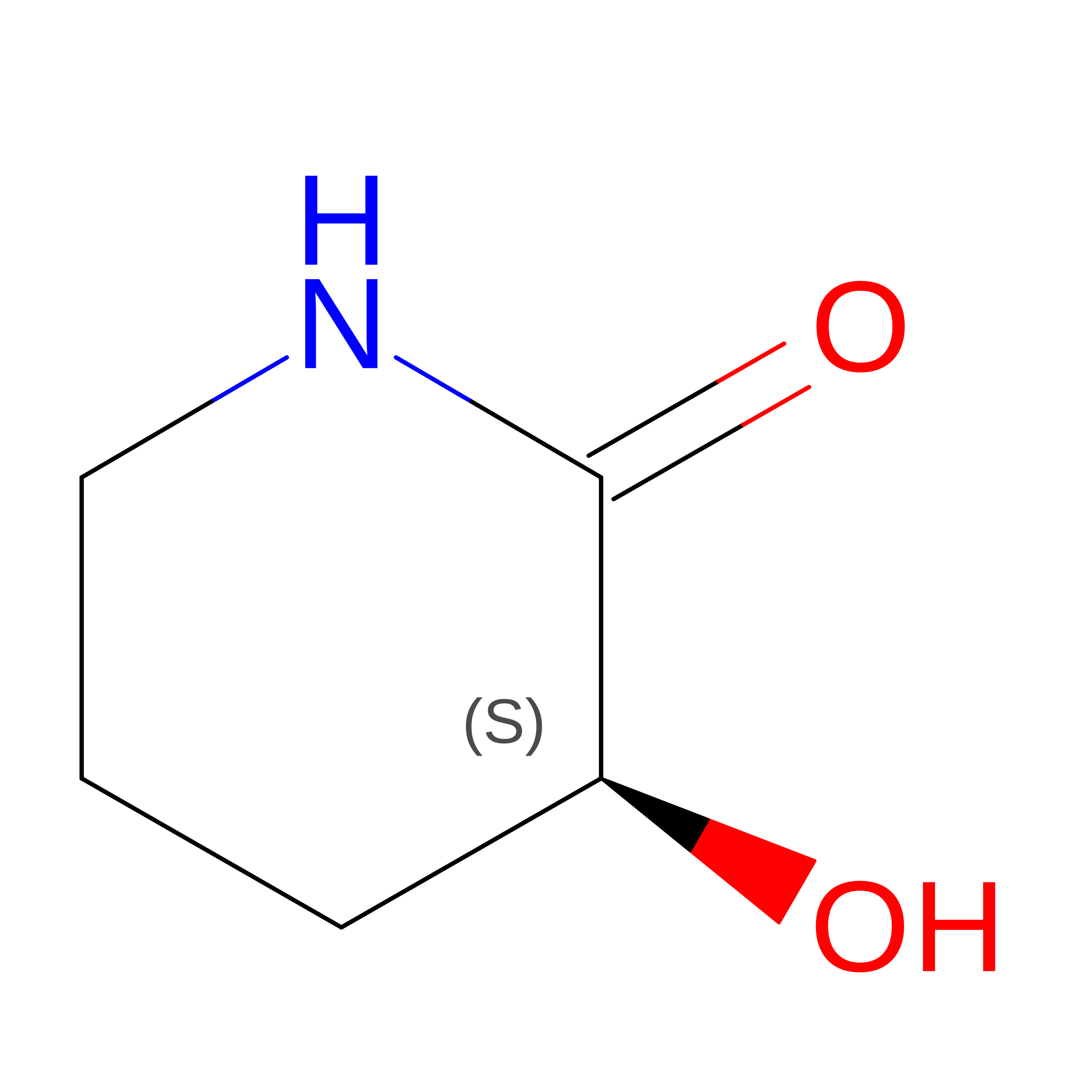
E [auth A],
I [auth A]
XYLOSE-DERIVED ISOFAGOMINE LACTAM
C5 H9 N O2
RYKLZUPYJFFNRR-BYPYZUCNSA-N
IMD

Query on IMD


Download:Ideal Coordinates CCD File
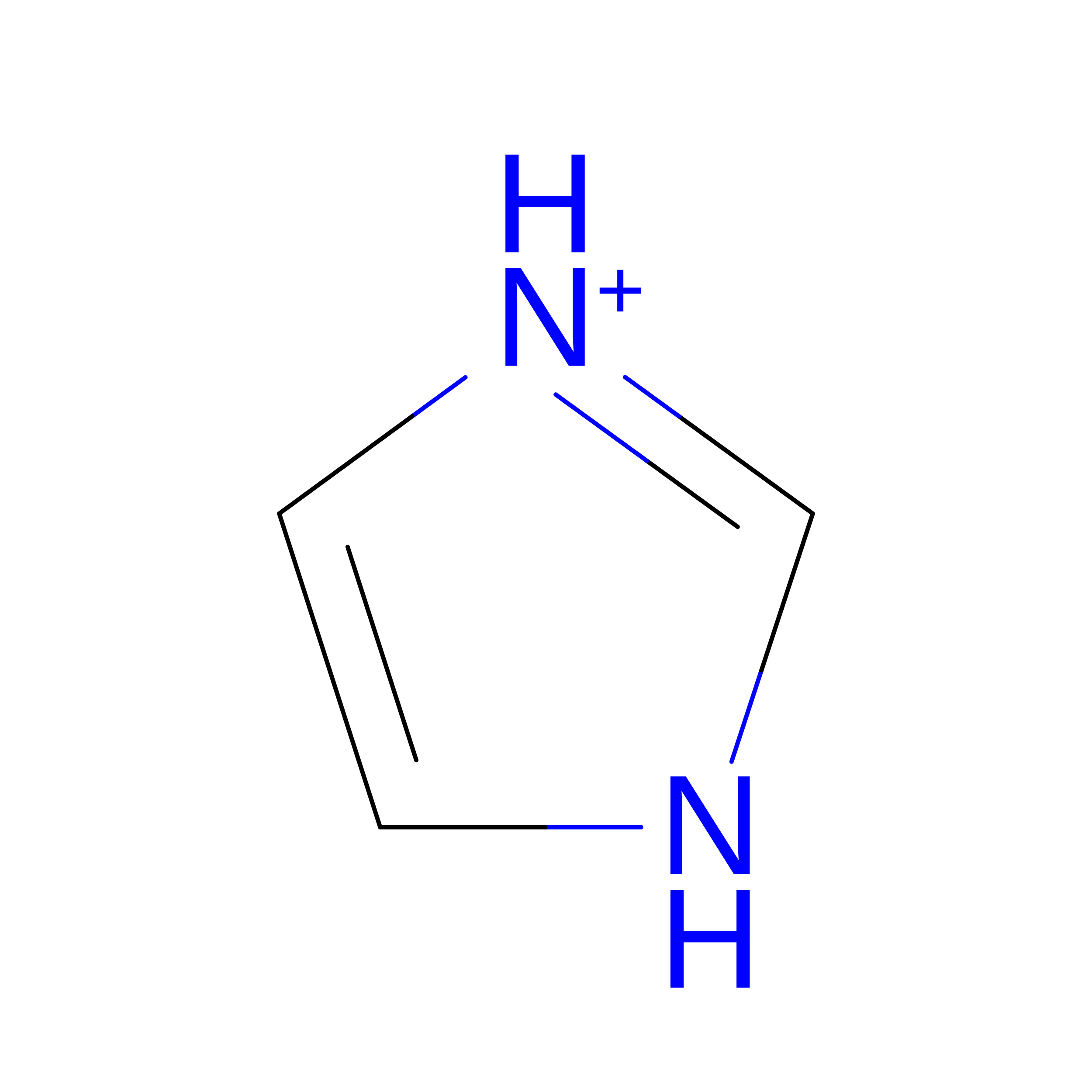
G [auth A],
H [auth A]
IMIDAZOLE
C3 H5 N2
RAXXELZNTBOGNW-UHFFFAOYSA-O
NA

Query on NA


Download:Ideal Coordinates CCD File
B [auth A],
C [auth A],
D [auth A],
K [auth A]
SODIUM ION
Na
FKNQFGJONOIPTF-UHFFFAOYSA-N

Experimental Data & Validation

Experimental Data

  • Method: X-RAY DIFFRACTION
  • Resolution: 1.05 Å
  • R-Value Free:  0.126 (Depositor), 0.136 (DCC) 
  • R-Value Work:  0.103 (Depositor), 0.119 (DCC) 
  • R-Value Observed: 0.104 (Depositor) 
Space Group: P 21 21 21
Unit Cell:
Length ( Å )Angle ( ˚ )
a = 65.814α = 90
b = 45.878β = 90
c = 85.737γ = 90
Software Package:
Software NamePurpose
REFMACrefinement
DENZOdata reduction
SCALEPACKdata scaling

Structure Validation

View Full Validation Report


Ligand Structure Quality Assessment 


Entry History 

Deposition Data

Revision History  (Full details and data files)

  • Version 1.0: 2003-04-08
    Type: Initial release
  • Version 1.1: 2011-05-08
    Changes: Version format compliance
  • Version 1.2: 2011-07-13
    Changes: Version format compliance
  • Version 2.0: 2020-07-29
    Type: Remediation
    Reason: Carbohydrate remediation
    Changes: Atomic model, Data collection, Derived calculations, Other, Structure summary
  • Version 2.1: 2023-12-13
    Changes: Data collection, Database references, Derived calculations, Refinement description, Structure summary
  • Version 2.2: 2024-11-13
    Changes: Structure summary