1OC0

plasminogen activator inhibitor-1 complex with somatomedin B domain of vitronectin


Experimental Data Snapshot

  • Method: X-RAY DIFFRACTION
  • Resolution: 2.28 Å
  • R-Value Free: 0.252 
  • R-Value Work: 0.196 
  • R-Value Observed: 0.198 

wwPDB Validation   3D Report Full Report


This is version 1.3 of the entry. See complete history


Literature

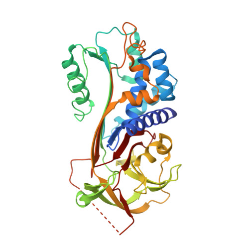
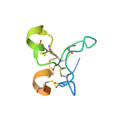
How Vitronectin Binds Pai-1 to Modulate Fibrinolysis and Cell Migration

Zhou, A.Huntington, J.A.Pannu, N.S.Carrell, R.W.Read, R.J.

(2003) Nat Struct Biol 10: 541

  • DOI: https://doi.org/10.1038/nsb943
  • Primary Citation of Related Structures:  
    1OC0

  • PubMed Abstract: 

    The interaction of the plasma protein vitronectin with plasminogen activator inhibitor-1 (PAI-1) is central to human health. Vitronectin binding extends the lifetime of active PAI-1, which controls hemostasis by inhibiting fibrinolysis and has also been implicated in angiogenesis. The PAI-1-vitronectin binding interaction also affects cell adhesion and motility. For these reasons, elevated PAI-1 activities are associated both with coronary thrombosis and with a poor prognosis in many cancers. Here we show the crystal structure at a resolution of 2.3 A of the complex of the somatomedin B domain of vitronectin with PAI-1. The structure of the complex explains how vitronectin binds to and stabilizes the active conformation of PAI-1. It also explains the tissue effects of PAI-1, as PAI-1 competes for and sterically blocks the interaction of vitronectin with cell surface receptors and integrins. Structural understanding of the essential biological roles of the interaction between PAI-1 and vitronectin opens the prospect of specifically designed blocking agents for the prevention of thrombosis and treatment of cancer.


  • Organizational Affiliation

    Department of Haematology, University of Cambridge, Cambridge Institute for Medical Research, Wellcome Trust/MRC Building, Hills Road, Cambridge CB2 2XY, UK.


Macromolecules
Find similar proteins by:  (by identity cutoff)  |  3D Structure
Entity ID: 1
MoleculeChains Sequence LengthOrganismDetailsImage
PLASMINOGEN ACTIVATOR INHIBITOR-1379Homo sapiensMutation(s): 4 
UniProt & NIH Common Fund Data Resources
Find proteins for P05121 (Homo sapiens)
Explore P05121 
Go to UniProtKB:  P05121
PHAROS:  P05121
GTEx:  ENSG00000106366 
Entity Groups  
Sequence Clusters30% Identity50% Identity70% Identity90% Identity95% Identity100% Identity
UniProt GroupP05121
Sequence Annotations
Expand
  • Reference Sequence
Find similar proteins by:  (by identity cutoff)  |  3D Structure
Entity ID: 2
MoleculeChains Sequence LengthOrganismDetailsImage
VITRONECTIN51Homo sapiensMutation(s): 0 
UniProt & NIH Common Fund Data Resources
Find proteins for P04004 (Homo sapiens)
Explore P04004 
Go to UniProtKB:  P04004
PHAROS:  P04004
GTEx:  ENSG00000109072 
Entity Groups  
Sequence Clusters30% Identity50% Identity70% Identity90% Identity95% Identity100% Identity
UniProt GroupP04004
Sequence Annotations
Expand
  • Reference Sequence
Experimental Data & Validation

Experimental Data

  • Method: X-RAY DIFFRACTION
  • Resolution: 2.28 Å
  • R-Value Free: 0.252 
  • R-Value Work: 0.196 
  • R-Value Observed: 0.198 
  • Space Group: P 21 21 21
Unit Cell:
Length ( Å )Angle ( ˚ )
a = 47.501α = 90
b = 90.087β = 90
c = 100.163γ = 90
Software Package:
Software NamePurpose
REFMACrefinement
DENZOdata reduction
SCALEPACKdata scaling
BEASTphasing

Structure Validation

View Full Validation Report


Entry History 

Deposition Data

Revision History  (Full details and data files)

  • Version 1.0: 2003-06-19
    Type: Initial release
  • Version 1.1: 2011-05-08
    Changes: Version format compliance
  • Version 1.2: 2011-07-13
    Changes: Version format compliance
  • Version 1.3: 2023-12-13
    Changes: Data collection, Database references, Other, Refinement description