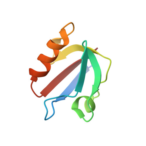
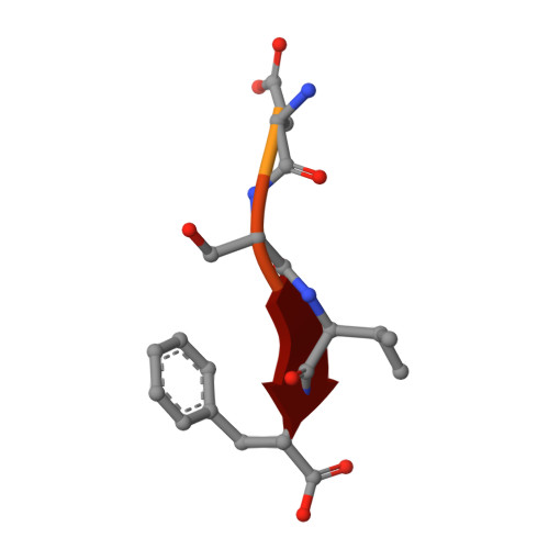
1OBX

Crystal structure of the complex of PDZ2 of syntenin with an interleukin 5 receptor alpha peptide.


Experimental Data Snapshot

  • Method: X-RAY DIFFRACTION
  • Resolution: 1.35 Å
  • R-Value Free: 0.212 
  • R-Value Work: 0.176 
  • R-Value Observed: 0.177 

wwPDB Validation   3D Report Full Report


This is version 1.3 of the entry. See complete history


Literature

Molecular Roots of Degenerate Specificity in Syntenin'S Pdz2 Domain: Reassessment of the Pdz Recognition Paradigm

Kang, B.S.Cooper, D.R.Devedjiev, Y.Derewenda, U.Derewenda, Z.S.

(2003) Structure 11: 845

  • DOI: 10.1016/s0969-2126(03)00125-4
  • Primary Citation of Related Structures:  
    1NTE, 1OBX, 1OBY, 1OBZ

  • PubMed Abstract: 
  • Crystal structures of the PDZ2 domain of the scaffolding protein syntenin, both unbound and in complexes with peptides derived from C termini of IL5 receptor (alpha chain) and syndecan, reveal the molecular roots of syntenin's degenerate specificity. Three distinct binding sites (S(0), S(-1), and S(-2)), with affinities for hydrophobic side chains, function in a combinatorial way: S(-1) and S(-2) act together to bind syndecan, while S(0) and S(-1) are involved in the binding of IL5Ralpha ...

    Crystal structures of the PDZ2 domain of the scaffolding protein syntenin, both unbound and in complexes with peptides derived from C termini of IL5 receptor (alpha chain) and syndecan, reveal the molecular roots of syntenin's degenerate specificity. Three distinct binding sites (S(0), S(-1), and S(-2)), with affinities for hydrophobic side chains, function in a combinatorial way: S(-1) and S(-2) act together to bind syndecan, while S(0) and S(-1) are involved in the binding of IL5Ralpha. Neither mode of interaction is consistent with the prior classification scheme, which defined the IL5Ralpha interaction as class I (-S/T-X-phi) and the syndecan interaction as class II (-phi-X-phi). These results, in conjunction with other emerging structural data on PDZ domains, call for a revision of their classification and of the existing model of their mechanism.


    Organizational Affiliation

    Department of Molecular Physiology and Biological Physics, The Cancer Center, University of Virginia, Charlottesville, VA 22908, USA.


Macromolecules
Find similar proteins by:  (by identity cutoff)  |  3D Structure
Entity ID: 1
MoleculeChainsSequence LengthOrganismDetailsImage
SYNTENIN 1A79Homo sapiensMutation(s): 0 
Gene Names: SDCBPMDA9SYCL
UniProt & NIH Common Fund Data Resources
Find proteins for O00560 (Homo sapiens)
Explore O00560 
Go to UniProtKB:  O00560
PHAROS:  O00560
Entity Groups  
Sequence Clusters30% Identity50% Identity70% Identity90% Identity95% Identity100% Identity
UniProt GroupO00560
Protein Feature View
Expand
  • Reference Sequence

Find similar proteins by:  Sequence   |   3D Structure  

Entity ID: 2
MoleculeChainsSequence LengthOrganismDetailsImage
INTERLEUKIN 5 RECEPTOR ALPHAB8Homo sapiensMutation(s): 0 
Gene Names: IL5RAIL5R
UniProt & NIH Common Fund Data Resources
Find proteins for Q01344 (Homo sapiens)
Explore Q01344 
Go to UniProtKB:  Q01344
PHAROS:  Q01344
Entity Groups  
UniProt GroupQ01344
Protein Feature View
Expand
  • Reference Sequence
Small Molecules
Ligands 2 Unique
IDChainsName / Formula / InChI Key2D Diagram3D Interactions
SO4
Query on SO4

Download Ideal Coordinates CCD File 
E [auth A],
F [auth A]
SULFATE ION
O4 S
QAOWNCQODCNURD-UHFFFAOYSA-L
 Ligand Interaction
CO
Query on CO

Download Ideal Coordinates CCD File 
C [auth A],
D [auth A]
COBALT (II) ION
Co
XLJKHNWPARRRJB-UHFFFAOYSA-N
 Ligand Interaction
Experimental Data & Validation

Experimental Data

  • Method: X-RAY DIFFRACTION
  • Resolution: 1.35 Å
  • R-Value Free: 0.212 
  • R-Value Work: 0.176 
  • R-Value Observed: 0.177 
  • Space Group: C 2 2 21
Unit Cell:
Length ( Å )Angle ( ˚ )
a = 53.723α = 90
b = 55.978β = 90
c = 51.094γ = 90
Software Package:
Software NamePurpose
REFMACrefinement
HKL-2000data reduction
HKL-2000data scaling
AMoREphasing

Structure Validation

View Full Validation Report


Entry History 

Deposition Data

Revision History  (Full details and data files)

  • Version 1.0: 2003-07-10
    Type: Initial release
  • Version 1.1: 2011-05-08
    Changes: Version format compliance
  • Version 1.2: 2011-07-13
    Changes: Version format compliance
  • Version 1.3: 2019-05-08
    Changes: Data collection, Experimental preparation, Other