1O8U

The 2 Angstrom Structure of 6-Oxo Camphor Hydrolase: New Structural Diversity in the Crotonase Superfamily


Experimental Data Snapshot

  • Method: X-RAY DIFFRACTION
  • Resolution: 2.00 Å
  • R-Value Free: 0.190 
  • R-Value Work: 0.147 
  • R-Value Observed: 0.149 

wwPDB Validation   3D Report Full Report


This is version 1.3 of the entry. See complete history


Literature

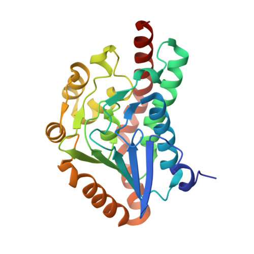
The 2 a Crystal Structure of 6-Oxo Camphor Hydrolase: New Structural Diversity in the Crotonase Superfamily

Whittingham, J.L.Turkenburg, J.P.Verma, C.S.Walsh, M.A.Grogan, G.

(2003) J Biol Chem 278: 1744

  • DOI: https://doi.org/10.1074/jbc.M211188200
  • Primary Citation of Related Structures:  
    1O8U

  • PubMed Abstract: 

    6-Oxo camphor hydrolase (OCH) is an enzyme of the crotonase superfamily that catalyzes carbon-carbon bond cleavage in bicyclic beta-diketones via a retro-Claisen reaction (Grogan, G., Roberts, G. A., Bougioukou, D., Turner, N. J., and Flitsch, S. L. (2001) J. Biol. Chem. 276, 12565-12572). The native structure of OCH has been solved at 2.0-A resolution with selenomethionine multiple wave anomalous dispersion and refined to a final R(free) of 19.0. The structure of OCH consists of a dimer of trimers that resembles the "parent" enzyme of the superfamily, enoyl-CoA hydratase. In contrast to enoyl-CoA hydratase, however, two octahedrally coordinated sodium atoms are found at the 3-fold axis of the hexamer of OCH, and the C-terminal helix of OCH does not form a discrete domain. Models of the substrate, 6-oxo camphor, and a proposed enolate intermediate in the putative active site suggest possible mechanistic roles for Glu-244, Asp-154, His-122, His-45, and His-145.


  • Organizational Affiliation

    York Structural Biology Laboratory, Department of Chemistry, University of York, Heslington, York, North Yorkshire YO10 5YW, United Kingdom.


Macromolecules
Find similar proteins by:  (by identity cutoff)  |  3D Structure
Entity ID: 1
MoleculeChains Sequence LengthOrganismDetailsImage
6-OXO CAMPHOR HYDROLASE
A, B, C, D, E
A, B, C, D, E, F
257Rhodococcus erythropolisMutation(s): 0 
UniProt
Find proteins for Q93TU6 (Rhodococcus sp)
Explore Q93TU6 
Go to UniProtKB:  Q93TU6
Entity Groups  
Sequence Clusters30% Identity50% Identity70% Identity90% Identity95% Identity100% Identity
UniProt GroupQ93TU6
Sequence Annotations
Expand
  • Reference Sequence
Small Molecules
Ligands 1 Unique
IDChains Name / Formula / InChI Key2D Diagram3D Interactions
NA
Query on NA

Download Ideal Coordinates CCD File 
G [auth A],
H [auth D]
SODIUM ION
Na
FKNQFGJONOIPTF-UHFFFAOYSA-N
Experimental Data & Validation

Experimental Data

  • Method: X-RAY DIFFRACTION
  • Resolution: 2.00 Å
  • R-Value Free: 0.190 
  • R-Value Work: 0.147 
  • R-Value Observed: 0.149 
  • Space Group: P 1 21 1
Unit Cell:
Length ( Å )Angle ( ˚ )
a = 78.95α = 90
b = 130.41β = 114.16
c = 81.32γ = 90
Software Package:
Software NamePurpose
REFMACrefinement
DENZOdata reduction
SCALEPACKdata scaling
SOLVEphasing

Structure Validation

View Full Validation Report


Entry History 

Deposition Data

Revision History  (Full details and data files)

  • Version 1.0: 2003-01-24
    Type: Initial release
  • Version 1.1: 2011-05-08
    Changes: Version format compliance
  • Version 1.2: 2011-07-13
    Changes: Version format compliance
  • Version 1.3: 2019-01-30
    Changes: Data collection, Experimental preparation