1O02 | pdb_00001o02

Human mitochondrial aldehyde dehydrogenase complexed with NADH in the presence of Mg2+


Experimental Data Snapshot

  • Method: X-RAY DIFFRACTION
  • Resolution: 1.90 Å
  • R-Value Free: 
    0.213 (Depositor), 0.213 (DCC) 
  • R-Value Work: 
    0.181 (Depositor), 0.180 (DCC) 
  • R-Value Observed: 
    0.185 (Depositor) 

Starting Model: experimental
View more details

wwPDB Validation 3D Report Full Report

Validation slider image for 1O02

Ligand Structure Quality Assessment 


This is version 1.5 of the entry. See complete history

Literature

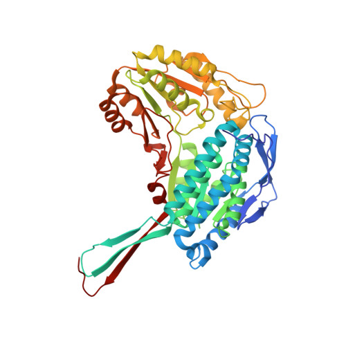
Coenzyme isomerization is integral to catalysis in aldehyde dehydrogenase

Perez-Miller, S.J.Hurley, T.D.

(2003) Biochemistry 42: 7100-7109

  • DOI: https://doi.org/10.1021/bi034182w
  • Primary Citation Related Structures: 
    1NZW, 1NZX, 1NZZ, 1O00, 1O01, 1O02, 1O04

  • PubMed Abstract: 

    Crystal structures of many enzymes in the aldehyde dehydrogenase superfamily determined in the presence of bound NAD(P)(+) have exhibited conformational flexibility for the nicotinamide half of the cofactor. This has been hypothesized to be important in catalysis because one conformation would block the second half of the reaction, but no firm evidence has been put forth which shows whether the oxidized and reduced cofactors preferentially occupy the two observed conformations. We present here two structures of the wild type and two structures of a Cys302Ser mutant of human mitochondrial aldehyde dehydrogenase in binary complexes with NAD(+) and NADH. These structures, including the Cys302Ser mutant in complex with NAD(+) at 1.4 A resolution and the wild-type enzyme in complex with NADH at 1.9 A resolution, provide strong evidence that bound NAD(+) prefers an extended conformation ideal for hydride transfer and bound NADH prefers a contracted conformation ideal for acyl-enzyme hydrolysis. Unique interactions between the cofactor and the Rossmann fold make isomerization possible while allowing the remainder of the active site complex to remain intact. In addition, these structures clarify the role of magnesium in activating the human class 2 enzyme. Our data suggest that the presence of magnesium may lead to selection of particular conformations and speed isomerization of the reduced cofactor following hydride transfer.


  • Organizational Affiliation
    • Program in Medical Biophysics and Department of Biochemistry and Molecular Biology, Indiana University School of Medicine, Indianapolis, Indiana 46202, USA.

Macromolecule Content 

  • Total Structure Weight: 443.14 kDa 
  • Atom Count: 34,432 
  • Modeled Residue Count: 3,952 
  • Deposited Residue Count: 4,000 
  • Unique protein chains: 1

Macromolecules

Find similar proteins by:|  3D Structure
Entity ID: 1
MoleculeChains  Sequence LengthOrganismDetailsImage
Aldehyde dehydrogenase
A, B, C, D, E
A, B, C, D, E, F, G, H
500Homo sapiensMutation(s): 0 
Gene Names: ALDH2 OR ALDM
EC: 1.2.1.3
UniProt & NIH Common Fund Data Resources
Find proteins for P05091 (Homo sapiens)
Explore P05091 
Go to UniProtKB:  P05091
PHAROS:  P05091
GTEx:  ENSG00000111275 
Entity Groups
Sequence Clusters30% Identity50% Identity70% Identity90% Identity95% Identity100% Identity
UniProt GroupP05091
Sequence Annotations
Expand
Reference Sequence

Small Molecules

Ligands 5 Unique
IDChains Name / Formula / InChI Key2D Diagram3D Interactions
NAI

Query on NAI


Download:Ideal Coordinates CCD File
DB [auth H]
FA [auth D]
LA [auth E]
N [auth A]
SA [auth F]
DB [auth H],
FA [auth D],
LA [auth E],
N [auth A],
SA [auth F],
T [auth B],
YA [auth G],
Z [auth C]
1,4-DIHYDRONICOTINAMIDE ADENINE DINUCLEOTIDE
C21 H29 N7 O14 P2
BOPGDPNILDQYTO-NNYOXOHSSA-N
EDO

Query on EDO


Download:Ideal Coordinates CCD File
CB [auth H]
EA [auth D]
KA [auth E]
M [auth A]
QA [auth F]
CB [auth H],
EA [auth D],
KA [auth E],
M [auth A],
QA [auth F],
RA [auth F],
S [auth B],
Y [auth C]
1,2-ETHANEDIOL
C2 H6 O2
LYCAIKOWRPUZTN-UHFFFAOYSA-N
GAI

Query on GAI


Download:Ideal Coordinates CCD File
BB [auth H]
CA [auth D]
DA [auth D]
IA [auth E]
JA [auth E]
BB [auth H],
CA [auth D],
DA [auth D],
IA [auth E],
JA [auth E],
K [auth A],
L [auth A],
OA [auth F],
PA [auth F],
Q [auth B],
R [auth B],
VA [auth G],
W [auth C],
WA [auth G],
X [auth C],
XA [auth G]
GUANIDINE
C H5 N3
ZRALSGWEFCBTJO-UHFFFAOYSA-N
MG

Query on MG


Download:Ideal Coordinates CCD File
AA [auth D]
GA [auth E]
I [auth A]
MA [auth F]
O [auth B]
AA [auth D],
GA [auth E],
I [auth A],
MA [auth F],
O [auth B],
TA [auth G],
U [auth C],
ZA [auth H]
MAGNESIUM ION
Mg
JLVVSXFLKOJNIY-UHFFFAOYSA-N
NA

Query on NA


Download:Ideal Coordinates CCD File
AB [auth H]
BA [auth D]
HA [auth E]
J [auth A]
NA [auth F]
AB [auth H],
BA [auth D],
HA [auth E],
J [auth A],
NA [auth F],
P [auth B],
UA [auth G],
V [auth C]
SODIUM ION
Na
FKNQFGJONOIPTF-UHFFFAOYSA-N

Experimental Data & Validation

Experimental Data

  • Method: X-RAY DIFFRACTION
  • Resolution: 1.90 Å
  • R-Value Free:  0.213 (Depositor), 0.213 (DCC) 
  • R-Value Work:  0.181 (Depositor), 0.180 (DCC) 
  • R-Value Observed: 0.185 (Depositor) 
Space Group: P 21 21 21
Unit Cell:
Length ( Å )Angle ( ˚ )
a = 141.492α = 90
b = 150.873β = 90
c = 177.195γ = 90
Software Package:
Software NamePurpose
CNSrefinement
DENZOdata reduction
SCALEPACKdata scaling
CNSphasing

Structure Validation

View Full Validation Report


Ligand Structure Quality Assessment 


Entry History 

Deposition Data

Revision History  (Full details and data files)

  • Version 1.0: 2003-06-24
    Type: Initial release
  • Version 1.1: 2008-04-29
    Changes: Version format compliance
  • Version 1.2: 2011-07-13
    Changes: Version format compliance
  • Version 1.3: 2013-10-30
    Changes: Non-polymer description
  • Version 1.4: 2019-02-27
    Changes: Advisory, Data collection
  • Version 1.5: 2023-08-16
    Changes: Data collection, Database references, Derived calculations, Refinement description