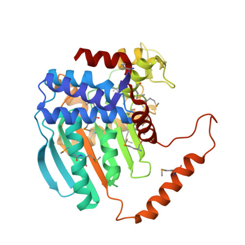
1NXU

CRYSTAL STRUCTURE OF E. COLI HYPOTHETICAL OXIDOREDUCTASE YIAK NORTHEAST STRUCTURAL GENOMICS CONSORTIUM TARGET ER82.


Experimental Data Snapshot

  • Method: X-RAY DIFFRACTION
  • Resolution: 1.80 Å
  • R-Value Free: 0.235 
  • R-Value Work: 0.192 
  • R-Value Observed: 0.192 

wwPDB Validation   3D Report Full Report


This is version 1.2 of the entry. See complete history


Literature

A Novel NAD-binding Protein Revealed by the Crystal Structure of 2,3-Diketo-L-gulonate Reductase (YiaK).

Forouhar, F.Lee, I.Benach, J.Kulkarni, K.Xiao, R.Acton, T.B.Montelione, G.T.Tong, L.

(2004) J Biol Chem 279: 13148-13155

  • DOI: https://doi.org/10.1074/jbc.M313580200
  • Primary Citation of Related Structures:  
    1NXU, 1S20

  • PubMed Abstract: 
  • Escherichia coli YiaK catalyzes the reduction of 2,3-diketo-L-gulonate in the presence of NADH. It belongs to a large family of oxidoreductases that is conserved in archaea, bacteria, and eukaryotes but shows no sequence homology to other proteins. We report here the crystal structures at up to 2 ...

    Escherichia coli YiaK catalyzes the reduction of 2,3-diketo-L-gulonate in the presence of NADH. It belongs to a large family of oxidoreductases that is conserved in archaea, bacteria, and eukaryotes but shows no sequence homology to other proteins. We report here the crystal structures at up to 2.0-A resolution of YiaK alone and in complex with NAD-tartrate. YiaK has a new polypeptide backbone fold and a novel mode of recognizing the NAD cofactor. In addition, NAD is bound in an unusual conformation, at the interface of a dimer of the enzyme. The crystallographic analysis unexpectedly revealed the binding of tartrate in the active site. Enzyme kinetics studies confirm that tartrate and the related D-malate are inhibitors of YiaK. In contrast to most other enzymes where substrate binding produces a more closed conformation, the binding of NAD-tartrate to YiaK produces a more open active site. The free enzyme conformation is incompatible with NAD binding. His(44) is likely the catalytic residue of the enzyme.


    Organizational Affiliation

    Department of Biological Sciences, Northeast Structural Genomics Consortium, Columbia University, New York, NY 10027, USA.


Macromolecules
Find similar proteins by:  (by identity cutoff)  |  3D Structure
Entity ID: 1
MoleculeChainsSequence LengthOrganismDetailsImage
Hypothetical oxidoreductase yiaK
A, B
333Escherichia coliMutation(s): 15 
Gene Names: YIAK OR B3575
EC: 1.1.1 (PDB Primary Data), 1.1.1.130 (UniProt)
UniProt
Find proteins for P37672 (Escherichia coli (strain K12))
Explore P37672 
Go to UniProtKB:  P37672
Entity Groups  
Sequence Clusters30% Identity50% Identity70% Identity90% Identity95% Identity100% Identity
UniProt GroupP37672
Protein Feature View
Expand
  • Reference Sequence
Small Molecules
Modified Residues  1 Unique
IDChainsTypeFormula2D DiagramParent
MSE
Query on MSE
A, B
L-PEPTIDE LINKINGC5 H11 N O2 SeMET
Experimental Data & Validation

Experimental Data

  • Method: X-RAY DIFFRACTION
  • Resolution: 1.80 Å
  • R-Value Free: 0.235 
  • R-Value Work: 0.192 
  • R-Value Observed: 0.192 
  • Space Group: P 1 21 1
Unit Cell:
Length ( Å )Angle ( ˚ )
a = 58.012α = 90
b = 51.229β = 103.78
c = 108.93γ = 90
Software Package:
Software NamePurpose
CNSrefinement
DENZOdata reduction
SCALEPACKdata scaling
SOLVEphasing

Structure Validation

View Full Validation Report


Entry History 

Revision History  (Full details and data files)

  • Version 1.0: 2003-03-11
    Type: Initial release
  • Version 1.1: 2008-04-29
    Changes: Version format compliance
  • Version 1.2: 2011-07-13
    Changes: Version format compliance