Crystallization and preliminary X-ray structural Studies of Hemoglobin A2 and Hemoglobin E, isolated from the blood samples of Beta-thalassemic patients
Dasgupta, J., Sen, U., Choudhury, D., Dutta, P., Chakrabarti, A., Chakrabarty, S.B., Chakrabarty, A., Dattagupta, J.K.(2004) Biochem Biophys Res Commun 303: 619-623
- PubMed: 12659864 Search on PubMed
- DOI: https://doi.org/10.1016/s0006-291x(03)00379-6
- Primary Citation Related Structures:
1NQP - PubMed Abstract:
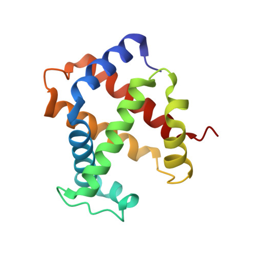
Hemoglobin A(2) (alpha(2)delta(2)), a minor (2-3%) component of circulating red blood cells, acts as an anti-sickling agent and its elevated concentration in beta-thalassemia is a useful clinical diagnostic. In beta-thalassemia major, where there is a failure of beta-chain production, HbA(2) acts as the predominant oxygen delivery mechanism. Hemoglobin E, is another common abnormal hemoglobin, caused by splice site mutation in exon 1 of beta globin gene, when combines with beta-thalassemia, causes severe microcytic anemia. The purification, crystallization, and preliminary structural studies of HbA(2) and HbE are reported here. HbA(2) and HbE are purified by cation exchange column chromatography in presence of KCN from the blood samples of individuals suffering from beta-thalassemia minor and E beta-thalassemia. X-ray diffraction data of HbA(2) and HbE were collected upto 2.1 and 1.73 A, respectively. HbA(2) crystallized in space group P2(1) with unit cell parameters a=54.33 A, b=83.73 A, c=62.87 A, and beta=99.80 degrees whereas HbE crystallized in space group P2(1)2(1)2(1) with unit cell parameters a=60.89 A, b=95.81 A, and c=99.08 A. Asymmetric unit in each case contains one Hb tetramer in R(2) state.
- Crystallography and Molecular Biology Division, Saha Institute of Nuclear Physics, 1/AF Bidhan Nagar, Kolkata 700064, India.
Organizational Affiliation: