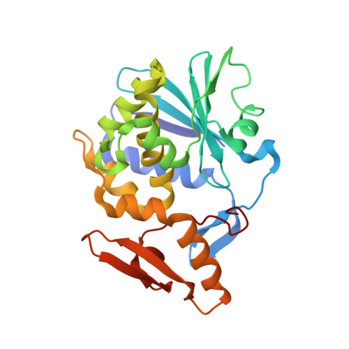
1NLI

Complex of [E160A-E189A] trichosanthin and adenine


Experimental Data Snapshot

  • Method: X-RAY DIFFRACTION
  • Resolution: 1.93 Å
  • R-Value Free: 0.208 
  • R-Value Work: 0.158 
  • R-Value Observed: 0.160 

Starting Model: experimental
View more details

wwPDB Validation   3D Report Full Report


This is version 1.4 of the entry. See complete history


Literature

Structural basis for the interaction of [E160A-E189A]-trichosanthin with adenine.

Shaw, P.C.Wong, K.B.Chan, D.S.Williams, R.L.

(2003) Toxicon 41: 575-581

  • DOI: https://doi.org/10.1016/s0041-0101(02)00387-2
  • Primary Citation of Related Structures:  
    1NLI

  • PubMed Abstract: 

    Trichosanthin is a ribosome-inactivating protein that cleaves specifically the N-glycosidic bond of A-4324 of 28S rRNA. Trichosanthin and its variant [E160A-E189A]-trichosanthin were found to bind an adenine base with a K(d) value of approximately 0.2mM. To determine how this doubly mutated variant of trichosanthin interacts with adenine, the co-crystal structure of [E160A-E189A]-trichosanthin and adenine was resolved to 0.193nm which revealed that the active site conformation of the doubly mutated variant is isomorphous to wild-type trichosanthin. Water molecules were found at locations corresponding to the eliminated side chain of Glu-160 and Glu-189. On the other hand, the adenine base interacted with [E160A-E189A]-trichosanthin in a manner similar to that in wild-type trichosanthin. Our structural analysis illustrates that Glu-160 and Glu-189 in trichosanthin do not play an important role in maintaining the active site conformation and binding adenine, an essential step for substrate-enzyme interaction. On the other hand, removal of two glutamate residues changed a large patch of negatively charged surface to a positive charge, which may account for the destabilization of the oxocarbenium-like transition-state and the significant decrease in ribosome-inactivating activity in [E160A-E189A]-trichosanthin.


  • Organizational Affiliation

    Department of Biochemistry, The Chinese University of Hong Kong, Shatin, NT, Hong Kong, People's Republic of China. pcshaw@cuhk.edu.hk


Macromolecules
Find similar proteins by:  (by identity cutoff)  |  3D Structure
Entity ID: 1
MoleculeChains Sequence LengthOrganismDetailsImage
Ribosome-inactivating protein alpha-trichosanthin248Trichosanthes kirilowiiMutation(s): 2 
Gene Names: Trichosanthin
EC: 3.2.2.22
UniProt
Find proteins for P09989 (Trichosanthes kirilowii)
Explore P09989 
Go to UniProtKB:  P09989
Entity Groups  
Sequence Clusters30% Identity50% Identity70% Identity90% Identity95% Identity100% Identity
UniProt GroupP09989
Sequence Annotations
Expand
  • Reference Sequence
Small Molecules
Ligands 1 Unique
IDChains Name / Formula / InChI Key2D Diagram3D Interactions
ADE
Query on ADE

Download Ideal Coordinates CCD File 
B [auth A]ADENINE
C5 H5 N5
GFFGJBXGBJISGV-UHFFFAOYSA-N
Binding Affinity Annotations 
IDSourceBinding Affinity
ADE PDBBind:  1NLI Kd: 2.60e+5 (nM) from 1 assay(s)
Experimental Data & Validation

Experimental Data

  • Method: X-RAY DIFFRACTION
  • Resolution: 1.93 Å
  • R-Value Free: 0.208 
  • R-Value Work: 0.158 
  • R-Value Observed: 0.160 
  • Space Group: P 21 21 21
Unit Cell:
Length ( Å )Angle ( ˚ )
a = 37.928α = 90
b = 75.299β = 90
c = 78.65γ = 90
Software Package:
Software NamePurpose
MOSFLMdata reduction
TRUNCATEdata reduction
AMoREphasing
REFMACrefinement
CCP4data scaling

Structure Validation

View Full Validation Report


Entry History 

Deposition Data

Revision History  (Full details and data files)

  • Version 1.0: 2003-01-21
    Type: Initial release
  • Version 1.1: 2008-04-29
    Changes: Version format compliance
  • Version 1.2: 2011-07-13
    Changes: Version format compliance
  • Version 1.3: 2021-10-27
    Changes: Database references, Derived calculations
  • Version 1.4: 2023-08-16
    Changes: Data collection, Refinement description