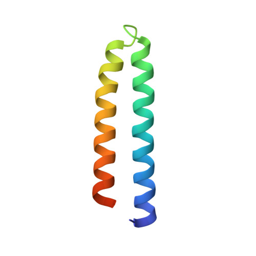
Structural parameters for proteins derived from the atomic resolution (1.09 A) structure of a designed variant of the ColE1 ROP protein.
Vlassi, M., Dauter, Z., Wilson, K.S., Kokkinidis, M.(1998) Acta Crystallogr D Biol Crystallogr 54: 1245-1260
- PubMed: 10089502
- DOI: https://doi.org/10.1107/s0907444998002492
- Primary Citation of Related Structures:
1NKD - PubMed Abstract:
The crystal structure of a designed variant of the ColE1 repressor of primer (ROP) protein has been refined with SHELXL93 to a resolution of 1.09 A. The final model with 510 non-H protein atoms, 576 H atoms in calculated positions and 114 water molecules converged to a standard R factor of 10% using unrestrained blocked full-matrix refinement. For all non-H atoms six-parameter anisotropic thermal parameters have been refined. The majority of atomic vibrations have a preferred orientation which is approximately perpendicular to the bundle axis; analysis with the TLS method [Schomaker & Trueblood (1968). Acta Cryst. B24, 63-77] showed a relatively good agreement between the individual atomic displacements and a rigid-body motion of the protein. Disordered residues with multiple conformations form clusters on the surface of the protein; six C-terminal residues have been omitted from the refined model due to disorder. Part of the solvent structure forms pentagonal or hexagonal clusters which bridge neighbouring protein molecules. Some water molecules are also conserved in wild-type ROP. The unrestrained blocked full-matrix least-squares refinement yielded reliable estimates of the standard deviations of the refined parameters. Comparison of these parameters with the stereochemical restraints used in various protein refinement programs showed statistically significant differences. These restraints should be adapted to the refinement of macromolecules by taking into account parameters determined from atomic resolution protein structures.
- National Centre for Scientific Research 'DEMOCRITOS', 15310 Ag. Paraskevi-Attikis, Athens, Greece.
Organizational Affiliation:


