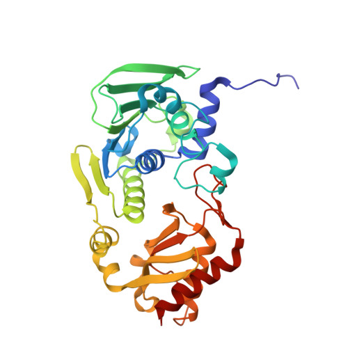
Monovalent cation dependence and preference of GHKL ATPases and kinases
Hu, X., Machius, M., Yang, W.(2003) FEBS Lett 544: 268-273
- PubMed: 12782329
- DOI: https://doi.org/10.1016/s0014-5793(03)00519-2
- Primary Citation of Related Structures:
1NHH, 1NHI, 1NHJ - PubMed Abstract:
The GHKL phosphotransferase superfamily, characterized by four sequence motifs that form the ATP-binding site, consists of the ATPase domains of type II DNA topoisomerases, Hsp90, and MutL, and bacterial and mitochondrial protein kinases. In addition to a magnesium ion, which is essential for catalysis, a potassium ion bound adjacent to the triphosphate moiety of ATP in a rat mitochondrial protein kinase, BCK (branched-chain alpha-ketoacid dehydrogenase kinase), has been shown to be indispensable for nucleotide binding and hydrolysis. Using X-ray crystallographic, biochemical, and genetic analyses, we find that the monovalent cation-binding site is conserved in MutL, but both Na(+) and K(+) support the MutL ATPase activity. When Ala100 of MutL is substituted by proline, mimicking the K(+)-binding environment in BCK, the mutant MutL protein becomes exclusively dependent on Na(+) for the ATPase activity. The coordination of this Na(+) ion is identical to that of the K(+) ion in BCK and involves four carbonyl oxygen atoms emanating from the hinges of the ATP lid and a non-bridging oxygen of the bound nucleotide. A similar monovalent cation-binding site is found in DNA gyrase with additional coordination by a serine side chain. The conserved and protein-specific monovalent cation-binding site is unique to the GHKL superfamily and probably essential for both ATPase and kinase activity. Dependence on different monovalent cations for catalysis may be exploited for future drug design specifically targeting each individual member of the GHKL superfamily.
- Laboratory of Molecular Biology, National Institute of Diabetes and Digestive and Kidney Diseases, National Institutes of Health, Bethesda, MD 20892, USA.
Organizational Affiliation:


