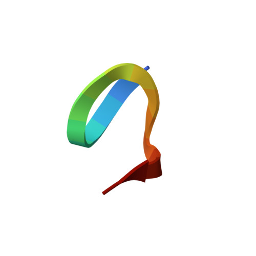
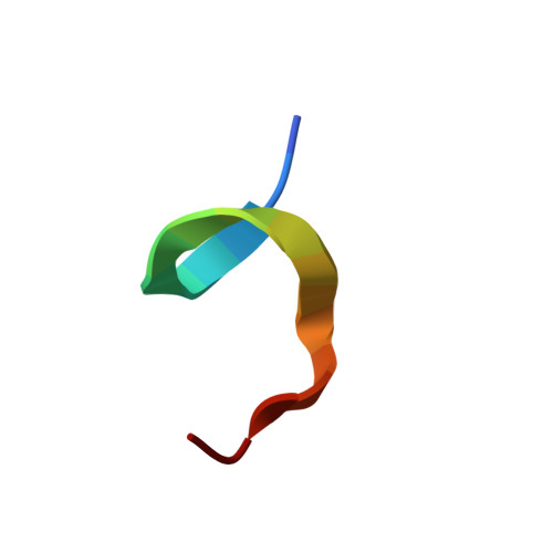
Structural characterization of a 39-residue synthetic peptide containing the two zinc binding domains from the HIV-1 p7 nucleocapsid protein by CD and NMR spectroscopy.
Omichinski, J.G., Clore, G.M., Sakaguchi, K., Appella, E., Gronenborn, A.M.(1991) FEBS Lett 292: 25-30
- PubMed: 1959614
- DOI: https://doi.org/10.1016/0014-5793(91)80825-n
- Primary Citation of Related Structures:
1NCP - PubMed Abstract:
A 39-residue peptide (p7-DF) containing the two zinc binding domains of the p7 nucleocapsid protein was prepared by solid-phase peptide synthesis. The solution structure of the peptide was characterized using circular dichroic and nuclear magnetic resonance spectroscopy in both the presence and absence of zinc ions. Circular dichroic spectroscopy indicates that the peptide exhibits a random coil conformation in the absence of zinc but appears to form an ordered structure in the presence of zinc. Two-dimensional nuclear magnetic resonance spectroscopy indicates that the two zinc binding domains within the peptide form stable, but independent, units upon the addition of 2 equivalents of ZnCl2 per equivalent of peptide. Structure calculations on the basis of nuclear Overhauser (NOE) data indicate that the two zinc binding domains have the same polypeptide fold within the errors of the coordinates (approximately 0.5 A for the backbone atoms, the zinc atoms and the coordinating cysteine and histidine ligands). The linker region (Arg17-Gly23) is characterized by a very limited number of sequential NOEs and the absence of any non-sequential NOEs suggest that this region of polypeptide chain is highly flexible. The latter coupled with the occurrence of a large number of basic residues (four out of seven) in the linker region suggests that it may serve to allow adaptable positioning of the nucleic acid recognition sequences within the protein.
- Laboratory of Chemical Physics, National Institute of Diabetes and Digestive and Kidney Diseases, National Institutes of Health, Bethesda, MD 20892.
Organizational Affiliation:


