Monitoring the Early Steps of Unfolding of Dicalcium and Mono-Ce(3+)-Substituted Forms of P43M Calbindin D(9k).
Jimenez, B., Poggi, L., Piccioli, M.(2003) Biochemistry 42: 13066-13073
- PubMed: 14596622 Search on PubMed
- DOI: https://doi.org/10.1021/bi034638+
- Primary Citation Related Structures:
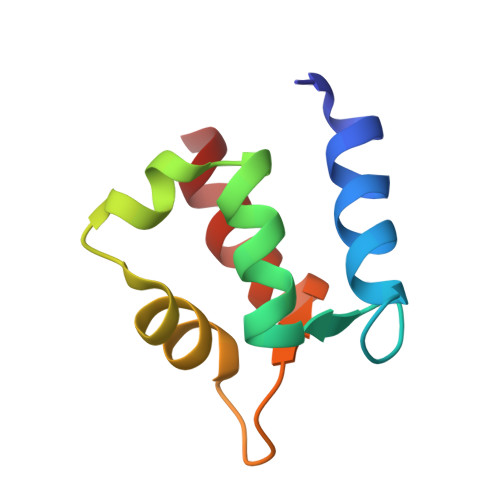
1N65 - PubMed Abstract:
Early steps of unfolding of P43M Calbindin D(9k) have been evaluated by NMR spectroscopy on the native dicalcium and on the paramagnetic monocerium-substituted derivative. Although at 2 M GdmHCl the protein core maintains its overall folding and structure, amide (15)N R(2) measurements and cross correlation rates between N-H dipole-dipole relaxation and (15)N CSA relaxation reveal a closer and stronger packing of the hydrophobic interactions in the protein as a response to the presence of denaturing agents in solution. A complete reorientation of the Met43 side chain toward the hydrophobic core is accomplished by the disappearance of the millisecond dynamics observed on the native form of Calbindin D(9k), while cross correlation rates provide evidence that the two-way hydrogen bond between Leu23 and Val61 is broken or substantially weakened. The substitution of the calcium ion in site II with the paramagnetic Ce(3+) ion allowed us to obtain a number of long-range nonconventional constraints, namely, pseudocontact shifts, which were used, together with the NOEs collected on the native state, to monitor subtle structural variations occurring in the non-native state of the protein. Although the average rmsd between the structures of native and non-native states is small (0.48 A), structural rearrangements could be reliably identified. Our results provide unprecedented information about the behavior of Calbindin D(9k) during the early steps of unfolding. Furthermore, they constitute strong evidence of the efficiency of paramagnetism-based constraints in monitoring subtle structural changes that are beyond the sensitivity of an approach based only on NOE.
- Departamento de Química Inorgánica, University of Valencia, Dr. Moliner, 50, 46100 Burjasot, Valencia, Spain.
Organizational Affiliation: