1N61

Crystal Structure of the Cu,Mo-CO Dehydrogenase (CODH); Dithionite reduced state


Experimental Data Snapshot

  • Method: X-RAY DIFFRACTION
  • Resolution: 1.30 Å
  • R-Value Free: 0.184 
  • R-Value Work: 0.142 
  • R-Value Observed: 0.145 

wwPDB Validation   3D Report Full Report


This is version 1.3 of the entry. See complete history


Literature

Catalysis at a dinuclear [CuSMo(=O)OH] cluster in a CO dehydrogenase resolved at 1.1-A resolution

Dobbek, H.Gremer, L.Kiefersauer, R.Huber, R.Meyer, O.

(2002) Proc Natl Acad Sci U S A 99: 15971-15976

  • DOI: https://doi.org/10.1073/pnas.212640899
  • Primary Citation of Related Structures:  
    1N5W, 1N60, 1N61, 1N62, 1N63

  • PubMed Abstract: 

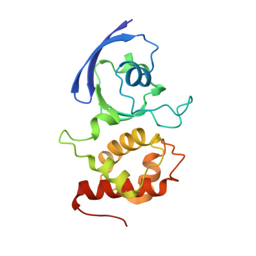
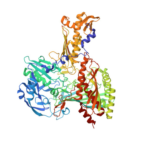
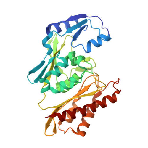
    The CO dehydrogenase of the eubacterium Oligotropha carboxidovorans is a 277-kDa Mo- and Cu-containing iron-sulfur flavoprotein. Here, the enzyme's active site in the oxidized or reduced state, after inactivation with potassium cyanide or with n-butylisocyanide bound to the active site, has been reinvestigated by multiple wavelength anomalous dispersion measurements at atomic resolution, electron spin resonance spectroscopy, and chemical analyses. We present evidence for a dinuclear heterometal [CuSMoO)OH] cluster in the active site of the oxidized or reduced enzyme, which is prone to cyanolysis. The cluster is coordinated through interactions of the Mo with the dithiolate pyran ring of molybdopterin cytosine dinucleotide and of the Cu with the Sgamma of Cys-388, which is part of the active-site loop VAYRC(388)SFR. The previously reported active-site structure [Dobbek, H., Gremer, L., Meyer, O. & Huber, R. (1999) Proc. Natl. Acad. Sci. USA 96, 8884-8889] of an Mo with three oxygen ligands and an SeH-group bound to the Sgamma atom of Cys-388 could not be confirmed. The structure of CO dehydrogenase with the inhibitor n-butylisocyanide bound has led to a model for the catalytic mechanism of CO oxidation which involves a thiocarbonate-like intermediate state. The dinuclear [CuSMo(O)OH] cluster of CO dehydrogenase establishes a previously uncharacterized class of dinuclear molybdoenzymes containing the pterin cofactor.


  • Organizational Affiliation

    Abteilung Strukturforschung, Max-Planck-Institut für Biochemie, and Proteros Biostructures GmbH, D-82152 Martinsried, Germany Europe. dobbek@biochem.mpg.de


Macromolecules
Find similar proteins by:  (by identity cutoff)  |  3D Structure
Entity ID: 1
MoleculeChains Sequence LengthOrganismDetailsImage
Carbon monoxide dehydrogenase small chain
A, D
166Afipia carboxidovorans OM5Mutation(s): 0 
EC: 1.2.99.2
UniProt
Find proteins for P19921 (Afipia carboxidovorans (strain ATCC 49405 / DSM 1227 / KCTC 32145 / OM5))
Explore P19921 
Go to UniProtKB:  P19921
Entity Groups  
Sequence Clusters30% Identity50% Identity70% Identity90% Identity95% Identity100% Identity
UniProt GroupP19921
Sequence Annotations
Expand
  • Reference Sequence
Find similar proteins by:  (by identity cutoff)  |  3D Structure
Entity ID: 2
MoleculeChains Sequence LengthOrganismDetailsImage
Carbon monoxide dehydrogenase large chain
B, E
809Afipia carboxidovorans OM5Mutation(s): 0 
EC: 1.2.99.2
UniProt
Find proteins for P19919 (Afipia carboxidovorans (strain ATCC 49405 / DSM 1227 / KCTC 32145 / OM5))
Explore P19919 
Go to UniProtKB:  P19919
Entity Groups  
Sequence Clusters30% Identity50% Identity70% Identity90% Identity95% Identity100% Identity
UniProt GroupP19919
Sequence Annotations
Expand
  • Reference Sequence
Find similar proteins by:  (by identity cutoff)  |  3D Structure
Entity ID: 3
MoleculeChains Sequence LengthOrganismDetailsImage
Carbon monoxide dehydrogenase medium chain
C, F
288Afipia carboxidovorans OM5Mutation(s): 0 
EC: 1.2.99.2
UniProt
Find proteins for P19920 (Afipia carboxidovorans (strain ATCC 49405 / DSM 1227 / KCTC 32145 / OM5))
Explore P19920 
Go to UniProtKB:  P19920
Entity Groups  
Sequence Clusters30% Identity50% Identity70% Identity90% Identity95% Identity100% Identity
UniProt GroupP19920
Sequence Annotations
Expand
  • Reference Sequence
Small Molecules
Ligands 5 Unique
IDChains Name / Formula / InChI Key2D Diagram3D Interactions
FAD
Query on FAD

Download Ideal Coordinates CCD File 
L [auth C],
Q [auth F]
FLAVIN-ADENINE DINUCLEOTIDE
C27 H33 N9 O15 P2
VWWQXMAJTJZDQX-UYBVJOGSSA-N
MCN
Query on MCN

Download Ideal Coordinates CCD File 
K [auth B],
P [auth E]
PTERIN CYTOSINE DINUCLEOTIDE
C19 H22 N8 O13 P2 S2
RBWYFPNWTRZKKZ-LOIMWUFNSA-N
CUN
Query on CUN

Download Ideal Coordinates CCD File 
J [auth B],
O [auth E]
CU(I)-S-MO(IV)(=O)OH CLUSTER
Cu H Mo O2 S
GIYZUGIRDQLYSY-UHFFFAOYSA-M
FES
Query on FES

Download Ideal Coordinates CCD File 
H [auth A],
I [auth A],
M [auth D],
N [auth D]
FE2/S2 (INORGANIC) CLUSTER
Fe2 S2
NIXDOXVAJZFRNF-UHFFFAOYSA-N
PO4
Query on PO4

Download Ideal Coordinates CCD File 
G [auth A]PHOSPHATE ION
O4 P
NBIIXXVUZAFLBC-UHFFFAOYSA-K
Experimental Data & Validation

Experimental Data

  • Method: X-RAY DIFFRACTION
  • Resolution: 1.30 Å
  • R-Value Free: 0.184 
  • R-Value Work: 0.142 
  • R-Value Observed: 0.145 
  • Space Group: P 21 21 21
Unit Cell:
Length ( Å )Angle ( ˚ )
a = 119.162α = 90
b = 131.375β = 90
c = 160.318γ = 90
Software Package:
Software NamePurpose
DENZOdata reduction
SCALEPACKdata scaling
CNSrefinement
REFMACrefinement
CNSphasing

Structure Validation

View Full Validation Report


Entry History 

Deposition Data

Revision History  (Full details and data files)

  • Version 1.0: 2002-12-18
    Type: Initial release
  • Version 1.1: 2008-04-28
    Changes: Version format compliance
  • Version 1.2: 2011-07-13
    Changes: Source and taxonomy, Version format compliance
  • Version 1.3: 2024-03-13
    Changes: Data collection, Database references, Derived calculations