1N3C

Structural and biochemical exploration of a critical amino acid in human 8-oxoguanine glycosylase


Experimental Data Snapshot

  • Method: X-RAY DIFFRACTION
  • Resolution: 2.70 Å
  • R-Value Free: 0.273 
  • R-Value Work: 0.225 
  • R-Value Observed: 0.225 

wwPDB Validation   3D Report Full Report


This is version 1.4 of the entry. See complete history


Literature

Structural and biochemical exploration of a critical amino acid in human 8-oxoguanine glycosylase

Norman, D.P.Chung, S.J.Verdine, G.L.

(2003) Biochemistry 42: 1564-1572

  • DOI: https://doi.org/10.1021/bi026823d
  • Primary Citation of Related Structures:  
    1N39, 1N3A, 1N3C

  • PubMed Abstract: 

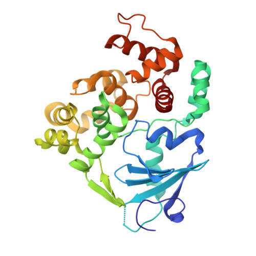
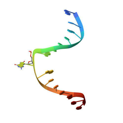
    Members of the HhH-GPD superfamily of DNA glycosylases are responsible for the recognition and removal of damaged nucleobases from DNA. The hallmark of these proteins is a motif comprising a helix-hairpin-helix followed by a Gly/Pro-rich loop and terminating in an invariant, catalytically essential aspartic acid residue. In this study, we have probed the role of this Asp in human 8-oxoguanine DNA glycosylase (hOgg1) by mutating it to Asn (D268N), Glu (D268E), and Gln (D268Q). We show that this aspartate plays a dual role, acting both as an N-terminal alpha-helix cap and as a critical residue for catalysis of both base excision and DNA strand cleavage by hOgg1. Mutation of this residue to asparagine, another helix-capping residue, preserves stability of the protein while drastically reducing enzymatic activity. A crystal structure of this mutant is the first to reveal the active site nucleophile Lys249 in the presence of lesion-containing DNA; this structure offers a tantalizing suggestion that base excision may occur by cleavage of the glycosidic bond and then attachment of Lys249. Mutation of the aspartic acid to glutamine and glutamic acid destabilizes the protein fold to a significant extent but, surprisingly, preserves catalytic activity. Crystal structures of these mutants complexed with an unreactive abasic site in DNA reveal these residues to adopt a sterically disfavored helix-capping conformation.


  • Organizational Affiliation

    Department of Chemistry and Chemical Biology, Harvard University, 12 Oxford Street, Cambridge, Massachusetts 02138, USA.


Macromolecules

Find similar proteins by:  (by identity cutoff)  |  3D Structure
Entity ID: 3
MoleculeChains Sequence LengthOrganismDetailsImage
N-glycosylase/DNA lyaseC [auth A]317Homo sapiensMutation(s): 1 
Gene Names: OGG1
UniProt & NIH Common Fund Data Resources
Find proteins for O15527 (Homo sapiens)
Explore O15527 
Go to UniProtKB:  O15527
PHAROS:  O15527
GTEx:  ENSG00000114026 
Entity Groups  
Sequence Clusters30% Identity50% Identity70% Identity90% Identity95% Identity100% Identity
UniProt GroupO15527
Sequence Annotations
Expand
  • Reference Sequence

Find similar nucleic acids by:  Sequence   |   3D Structure  

Entity ID: 1
MoleculeChains LengthOrganismImage
DNA complement strandA [auth B]15N/A
Sequence Annotations
Expand
  • Reference Sequence

Find similar nucleic acids by:  Sequence   |   3D Structure  

Entity ID: 2
MoleculeChains LengthOrganismImage
8-oxoG-containing DNAB [auth C]15N/A
Sequence Annotations
Expand
  • Reference Sequence
Small Molecules
Ligands 1 Unique
IDChains Name / Formula / InChI Key2D Diagram3D Interactions
CA
Query on CA

Download Ideal Coordinates CCD File 
D [auth C]CALCIUM ION
Ca
BHPQYMZQTOCNFJ-UHFFFAOYSA-N
Binding Affinity Annotations 
IDSourceBinding Affinity
DNA PDBBind:  1N3C Kd: 700 (nM) from 1 assay(s)
Experimental Data & Validation

Experimental Data

  • Method: X-RAY DIFFRACTION
  • Resolution: 2.70 Å
  • R-Value Free: 0.273 
  • R-Value Work: 0.225 
  • R-Value Observed: 0.225 
  • Space Group: P 65 2 2
Unit Cell:
Length ( Å )Angle ( ˚ )
a = 92.255α = 90
b = 92.255β = 90
c = 210.6γ = 120
Software Package:
Software NamePurpose
HKL-2000data collection
SCALEPACKdata scaling
CNSrefinement
HKL-2000data reduction
CNSphasing

Structure Validation

View Full Validation Report


Entry History 

Deposition Data

Revision History  (Full details and data files)

  • Version 1.0: 2003-03-04
    Type: Initial release
  • Version 1.1: 2008-04-28
    Changes: Version format compliance
  • Version 1.2: 2011-07-13
    Changes: Version format compliance
  • Version 1.3: 2021-10-27
    Changes: Database references, Derived calculations
  • Version 1.4: 2024-02-14
    Changes: Data collection, Refinement description