1MZN

CRYSTAL STRUCTURE at 1.9 ANGSTROEMS RESOLUTION OF THE HOMODIMER OF HUMAN RXR ALPHA LIGAND BINDING DOMAIN BOUND TO THE SYNTHETIC AGONIST COMPOUND BMS 649 AND A COACTIVATOR PEPTIDE


Experimental Data Snapshot

  • Method: X-RAY DIFFRACTION
  • Resolution: 1.90 Å
  • R-Value Free: 0.232 
  • R-Value Work: 0.195 
  • R-Value Observed: 0.195 

wwPDB Validation   3D Report Full Report


This is version 1.3 of the entry. See complete history


Literature

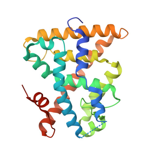
Molecular Recognition of Agonist Ligands by RXRs

Egea, P.F.Mitschler, A.Moras, D.

(2002) Mol Endocrinol 16: 987-997

  • DOI: 10.1210/mend.16.5.0823
  • Primary Citation of Related Structures:  
    1MV9, 1MVC, 1MZN

  • PubMed Abstract: 
  • The nuclear receptor RXR is an obligate partner in many signal transduction pathways. We report the high-resolution structures of two complexes of the human RXRalpha ligand-binding domain specifically bound to two different and chemically unrelated agonist compounds: docosa hexaenoic acid, a natural derivative of eicosanoic acid, present in mammalian cells and recently identified as a potential endogenous RXR ligand in the mouse brain, and the synthetic ligand BMS 649 ...

    The nuclear receptor RXR is an obligate partner in many signal transduction pathways. We report the high-resolution structures of two complexes of the human RXRalpha ligand-binding domain specifically bound to two different and chemically unrelated agonist compounds: docosa hexaenoic acid, a natural derivative of eicosanoic acid, present in mammalian cells and recently identified as a potential endogenous RXR ligand in the mouse brain, and the synthetic ligand BMS 649. In both structures the RXR-ligand-binding domain forms homodimers and exhibits the active conformation previously observed with 9-cis-RA. Analysis of the differences in ligand-protein contacts (predominantly van der Waals forces) and binding cavity geometries and volumes for the several agonist-bound RXR structures clarifies the structural features important for ligand recognition. The L-shaped ligand-binding pocket adapts to the diverse ligands, especially at the level of residue N306, which might thus constitute a new target for drug-design. Despite its highest affinity 9-cis-RA displays the lowest number of ligand-protein contacts. These structural results support the idea that docosa hexaenoic acid and related fatty acids could be natural agonists of RXRs and question the real nature of the endogenous ligand(s) in mammalian cells.


    Related Citations: 
    • Crystal structure of the human RXRalpha ligand-binding domain bound to its natural ligand: 9-cis retinoic acid
      Egea, P.F., Mitschler, A., Rochel, N., Ruff, M., Chambon, P., Moras, D.
      (2000) EMBO J 19: 2592

    Organizational Affiliation

    Laboratoire de Biologie et Génomique Structurales, Université Louis Pasteur, Parc d'Innovation BP163, 67404 Illkirch cedex, France.


Macromolecules
Find similar proteins by:  (by identity cutoff)  |  3D Structure
Entity ID: 1
MoleculeChainsSequence LengthOrganismDetailsImage
RXR retinoid X receptorA, C, E, G240Homo sapiensMutation(s): 0 
Gene Names: RXRANR2B1
UniProt & NIH Common Fund Data Resources
Find proteins for P19793 (Homo sapiens)
Explore P19793 
Go to UniProtKB:  P19793
PHAROS:  P19793
Entity Groups  
Sequence Clusters30% Identity50% Identity70% Identity90% Identity95% Identity100% Identity
UniProt GroupP19793
Protein Feature View
Expand
  • Reference Sequence

Find similar proteins by:  Sequence   |   3D Structure  

Entity ID: 2
MoleculeChainsSequence LengthOrganismDetailsImage
Nuclear receptor coactivator 2B, D, F, H13synthetic constructMutation(s): 0 
UniProt & NIH Common Fund Data Resources
Find proteins for Q15596 (Homo sapiens)
Explore Q15596 
Go to UniProtKB:  Q15596
PHAROS:  Q15596
Entity Groups  
Sequence Clusters30% Identity50% Identity70% Identity90% Identity95% Identity100% Identity
UniProt GroupQ15596
Protein Feature View
Expand
  • Reference Sequence
Small Molecules
Ligands 1 Unique
IDChainsName / Formula / InChI Key2D Diagram3D Interactions
BM6
Query on BM6

Download Ideal Coordinates CCD File 
I [auth A],
J [auth C],
K [auth E],
L [auth G]
4-[2-(5,5,8,8-TETRAMETHYL-5,6,7,8-TETRAHYDRO-NAPHTHALEN-2-YL)-[1,3]DIOXOLAN-2-YL]-BENZOIC ACID
C24 H27 O4
ZZUKALQMHNSWTK-UHFFFAOYSA-M
 Ligand Interaction
Experimental Data & Validation

Experimental Data

  • Method: X-RAY DIFFRACTION
  • Resolution: 1.90 Å
  • R-Value Free: 0.232 
  • R-Value Work: 0.195 
  • R-Value Observed: 0.195 
  • Space Group: P 1
Unit Cell:
Length ( Å )Angle ( ˚ )
a = 47.119α = 109.97
b = 64.694β = 92.87
c = 94.784γ = 89.99
Software Package:
Software NamePurpose
DENZOdata reduction
SCALEPACKdata scaling
AMoREphasing
CNSrefinement

Structure Validation

View Full Validation Report


Entry History 

Deposition Data

Revision History  (Full details and data files)

  • Version 1.0: 2002-10-23
    Type: Initial release
  • Version 1.1: 2008-04-28
    Changes: Version format compliance
  • Version 1.2: 2011-07-13
    Changes: Version format compliance
  • Version 1.3: 2021-08-04
    Changes: Data collection, Derived calculations, Refinement description, Source and taxonomy