1MZ9

Storage function of COMP:the crystal structure of the coiled-coil domain in complex with vitamin D3


Experimental Data Snapshot

  • Method: X-RAY DIFFRACTION
  • Resolution: 1.70 Å
  • R-Value Free: 0.254 
  • R-Value Work: 0.231 
  • R-Value Observed: 0.231 

wwPDB Validation   3D Report Full Report


This is version 1.3 of the entry. See complete history


Literature

Storage function of cartilage oligomeric matrix protein: the crystal structure of the coiled-coil domain in complex with vitamin D(3).

Ozbek, S.Engel, J.Stetefeld, J.

(2002) EMBO J 21: 5960-5968

  • DOI: https://doi.org/10.1093/emboj/cdf628
  • Primary Citation of Related Structures:  
    1MZ9

  • PubMed Abstract: 

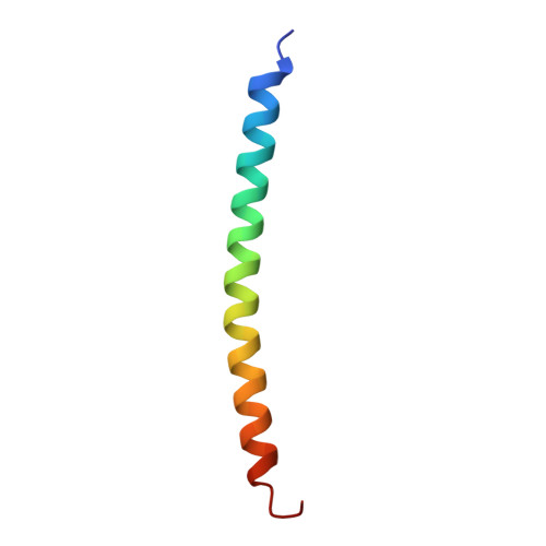
    The five-stranded coiled-coil domain of cartilage oligomeric matrix protein (COMPcc) forms a continuous axial pore with binding capacities for hydrophobic compounds, including prominent cell signalling molecules. Here, we report the X-ray structure of the COMPcc domain in complex with vitamin D(3) at 1.7 A resolution. The COMPcc pentamer harbours two molecules of the steroid hormone precursor in a planar s-trans conformation of the conjugated triene, with the aliphatic tails lying along the molecule axis. A hydrophilic ring of five Gln54 side chains divides the channel into two hydrophobic compartments in which the bound vitamin D(3) pair is fixed in a head-to-head orientation. Vitamin D(3) binding induces a volumetric increase of the cavities of approximately 30% while the main chain distances of the pentamer are retained. This adaptation to the bulky ring systems of the ligands is accomplished by a rotamer re-orientation of beta-branched side chains that form the knobs into holes of the coiled-coil structure. Compared with binding of vitamin D and retinoic acid by their classical receptors, COMP exerts a distinct mechanism of interaction mainly defined by the pattern of hydrophobic core residues.


  • Organizational Affiliation

    Department of Biophysical Chemistry, Biozentrum, University of Basel, Klingelbergstrasse 70, CH-4056 Basel, Switzerland. Suat.Oezbek@unibas.ch


Macromolecules
Find similar proteins by:  (by identity cutoff)  |  3D Structure
Entity ID: 1
MoleculeChains Sequence LengthOrganismDetailsImage
cartilage oligomeric matrix protein
A, B, C, D, E
45Mus musculusMutation(s): 0 
UniProt
Find proteins for Q9R0G6 (Mus musculus)
Explore Q9R0G6 
Go to UniProtKB:  Q9R0G6
Entity Groups  
Sequence Clusters30% Identity50% Identity70% Identity90% Identity95% Identity100% Identity
UniProt GroupQ9R0G6
Sequence Annotations
Expand
  • Reference Sequence
Small Molecules
Ligands 1 Unique
IDChains Name / Formula / InChI Key2D Diagram3D Interactions
VDY
Query on VDY

Download Ideal Coordinates CCD File 
F [auth A],
G [auth D]
3-{2-[1-(5-HYDROXY-1,5-DIMETHYL-HEXYL)-7A-METHYL-OCTAHYDRO-INDEN-4-YLIDENE]-ETHYLIDENE}-4-METHYLENE-CYCLOHEXANOL
C27 H44 O2
JWUBBDSIWDLEOM-DTOXIADCSA-N
Experimental Data & Validation

Experimental Data

  • Method: X-RAY DIFFRACTION
  • Resolution: 1.70 Å
  • R-Value Free: 0.254 
  • R-Value Work: 0.231 
  • R-Value Observed: 0.231 
  • Space Group: P 1 21 1
Unit Cell:
Length ( Å )Angle ( ˚ )
a = 38.295α = 90
b = 49.289β = 103.87
c = 55.26γ = 90
Software Package:
Software NamePurpose
MOSFLMdata reduction
SCALAdata scaling
AMoREphasing
CNSrefinement
CCP4data scaling

Structure Validation

View Full Validation Report


Entry History 

Deposition Data

Revision History  (Full details and data files)

  • Version 1.0: 2003-01-28
    Type: Initial release
  • Version 1.1: 2008-04-28
    Changes: Version format compliance
  • Version 1.2: 2011-07-13
    Changes: Version format compliance
  • Version 1.3: 2022-12-21
    Changes: Data collection, Database references, Derived calculations