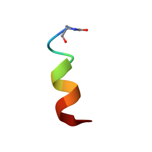
Solution structure of the tachykinin Peptide eledoisin
Grace, R.C., Chandrashekar, I.R., Cowsik, S.M.(2003) Biophys J 84: 655-664
- PubMed: 12524318 Search on PubMedSearch on PubMed Central
- DOI: https://doi.org/10.1016/S0006-3495(03)74885-1
- Primary Citation Related Structures:
1MXQ - PubMed Abstract:
Both the aqueous and the lipid-induced structure of eledoisin, an undecapeptide of mollusk origin, have been studied by two-dimensional proton nuclear magnetic resonance spectroscopy and distance geometry calculations. Unambiguous nuclear magnetic resonance assignments of protons have been made with the aid of correlation spectroscopy experiments and nuclear Overhauser effect spectroscopy experiments. The distance constraints obtained from the nuclear magnetic resonance data have been utilized in a distance geometry algorithm to generate a family of structures, which have been refined using restrained energy minimization and dynamics. These data show that, while in water and dimethyl sulfoxide, eledoisin prefers to be in an extended chain conformation, whereas in the presence of perdeuterated dodecylphosphocholine micelles, a membrane model system, helical conformation is induced in the central core and C-terminal region (K4-M11) of the peptide. N terminus, though less defined, also displays some degree of order and a possible turn structure. The conformation adopted by eledoisin in the presence of dodecylphosphocholine micelles is similar to the structural motif typical of neurokinin-2 selective agonists and with that reported for kassinin in hydrophobic environment.
- Post Graduate Department of Physics, Christ College, Bangalore, 560 029 India.
Organizational Affiliation: