1MQG

Crystal Structure of the GluR2 Ligand Binding Core (S1S2J) in Complex with Iodo-Willardiine at 2.15 Angstroms Resolution


Experimental Data Snapshot

  • Method: X-RAY DIFFRACTION
  • Resolution: 2.15 Å
  • R-Value Free: 0.233 
  • R-Value Work: 0.185 
  • R-Value Observed: 0.185 

wwPDB Validation   3D Report Full Report


This is version 1.3 of the entry. See complete history


Literature

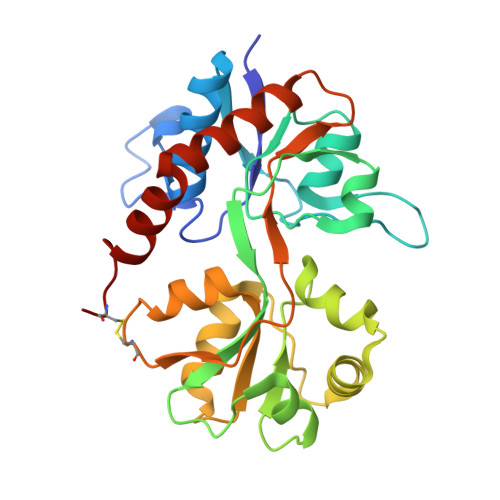
Structural basis for partial agonist action at ionotropic glutamate receptors

Jin, R.Banke, T.G.Mayer, M.L.Traynelis, S.F.Gouaux, E.

(2003) Nat Neurosci 6: 803-810

  • DOI: https://doi.org/10.1038/nn1091
  • Primary Citation of Related Structures:  
    1MQG, 1MQH, 1MQI, 1MQJ

  • PubMed Abstract: 

    An unresolved problem in understanding neurotransmitter receptor function concerns the mechanism(s) by which full and partial agonists elicit different amplitude responses at equal receptor occupancy. The widely held view of 'partial agonism' posits that resting and active states of the receptor are in equilibrium, and partial agonists simply do not shift the equilibrium toward the active state as efficaciously as full agonists. Here we report findings from crystallographic and electrophysiological studies of the mechanism of activation of an AMPA-subtype glutamate receptor ion channel. In these experiments, we used 5-substituted willardiines, a series of partial agonists that differ by only a single atom. Our results show that the GluR2 ligand-binding core can adopt a range of ligand-dependent conformational states, which in turn control the open probability of discrete subconductance states of the intact ion channel. Our findings thus provide a structure-based model of partial agonism.


  • Organizational Affiliation

    Department of Biochemistry and Molecular Biophysics, Columbia University, 650 West 168 Street, New York, New York 10032, USA.


Macromolecules
Find similar proteins by:  (by identity cutoff)  |  3D Structure
Entity ID: 1
MoleculeChains Sequence LengthOrganismDetailsImage
glutamate receptor 2
A, B
263Rattus norvegicusMutation(s): 0 
Gene Names: GluR-2 or GluR-B
UniProt
Find proteins for P19491 (Rattus norvegicus)
Explore P19491 
Go to UniProtKB:  P19491
Entity Groups  
Sequence Clusters30% Identity50% Identity70% Identity90% Identity95% Identity100% Identity
UniProt GroupP19491
Sequence Annotations
Expand
  • Reference Sequence
Small Molecules
Ligands 3 Unique
IDChains Name / Formula / InChI Key2D Diagram3D Interactions
IWD
Query on IWD

Download Ideal Coordinates CCD File 
D [auth A],
G [auth B]
2-AMINO-3-(5-IODO-2,4-DIOXO-3,4-DIHYDRO-2H-PYRIMIDIN-1-YL)-PROPIONIC ACID
C7 H8 I N3 O4
AXXYLTBQIQBTES-BYPYZUCNSA-N
SO4
Query on SO4

Download Ideal Coordinates CCD File 
C [auth A],
F [auth B]
SULFATE ION
O4 S
QAOWNCQODCNURD-UHFFFAOYSA-L
GOL
Query on GOL

Download Ideal Coordinates CCD File 
E [auth A]GLYCEROL
C3 H8 O3
PEDCQBHIVMGVHV-UHFFFAOYSA-N
Binding Affinity Annotations 
IDSourceBinding Affinity
IWD BindingDB:  1MQG Ki: min: 105, max: 976 (nM) from 5 assay(s)
Binding MOAD:  1MQG IC50: 520 (nM) from 1 assay(s)
PDBBind:  1MQG IC50: 520 (nM) from 1 assay(s)
Experimental Data & Validation

Experimental Data

  • Method: X-RAY DIFFRACTION
  • Resolution: 2.15 Å
  • R-Value Free: 0.233 
  • R-Value Work: 0.185 
  • R-Value Observed: 0.185 
  • Space Group: P 1 21 1
Unit Cell:
Length ( Å )Angle ( ˚ )
a = 48.006α = 90
b = 88.716β = 99.24
c = 58.907γ = 90
Software Package:
Software NamePurpose
DENZOdata reduction
SCALEPACKdata scaling
CNSrefinement
CNSphasing

Structure Validation

View Full Validation Report


Entry History 

Deposition Data

Revision History  (Full details and data files)

  • Version 1.0: 2003-08-05
    Type: Initial release
  • Version 1.1: 2008-04-28
    Changes: Version format compliance
  • Version 1.2: 2011-07-13
    Changes: Non-polymer description, Version format compliance
  • Version 1.3: 2017-05-10
    Changes: Derived calculations