1MNE | pdb_00001mne

TRUNCATED HEAD OF MYOSIN FROM DICTYOSTELIUM DISCOIDEUM COMPLEXED WITH MG-PYROPHOSPHATE


Experimental Data Snapshot

  • Method: X-RAY DIFFRACTION
  • Resolution: 2.70 Å

wwPDB Validation 3D Report Full Report

Validation slider image for 1MNE

This is version 1.3 of the entry. See complete history

Literature

X-ray structure of the magnesium(II)-pyrophosphate complex of the truncated head of Dictyostelium discoideum myosin to 2.7 A resolution.

Smith, C.A.Rayment, I.

(1995) Biochemistry 34: 8973-8981

  • DOI: https://doi.org/10.1021/bi00028a005
  • Primary Citation Related Structures: 
    1MNE

  • PubMed Abstract: 

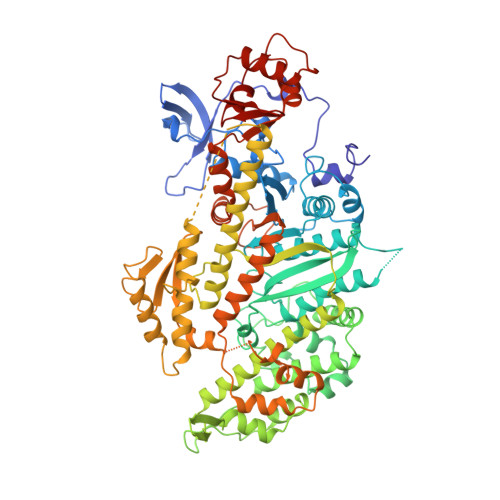
    The structure of the magnesium pyrophosphate complex of the truncated head of Dictyostelium myosin has been determined by molecular replacement at 2.7 A resolution and refined to a crystallographic R-factor of 16.0%. The crystals belong to the orthorhombic space group P2(1)2(1)2, where a = 105.2 A, b = 182.1 A, and c = 54.5 A. The conformation of the protein around the magnesium pyrophosphate is very similar to that seen when magnesium ADP-beryllium fluoride binds in the active site. The latter complex mimics the binding of ATP prior to hydrolysis. The pyrophosphate molecule occupies the beta- and gamma-phosphate sites, where the two phosphorus atoms are in the same positions as the beta-phosphate and the BeFx moiety of the beryllium fluoride-trapped ADP. The surrounding active site residues are almost perfectly superimposable in the two structures and the hydrogen-bonding interactions that the PPi makes with the protein are essentially identical. The similarity between the MgPPi and MgADP.BeFx complex with S1Dc suggests that the conformational change, which occurs when ATP binds to actomyosin and which reduces the affinity of myosin for actin, is caused by the binding of the gamma- and beta-phosphate groups of the nucleotide. This then implies that the role of the remainder of the substrate is to increase the binding affinity for myosin and thus to drive the equilibrium toward dissociation of myosin from actin.


  • Organizational Affiliation
    • Institute for Enzyme Research, University of Wisconsin, Madison 53705, USA.

Macromolecule Content 

  • Total Structure Weight: 86.95 kDa 
  • Atom Count: 6,027 
  • Modeled Residue Count: 742 
  • Deposited Residue Count: 762 
  • Unique protein chains: 1

Macromolecules

Find similar proteins by:|  3D Structure
Entity ID: 1
MoleculeChains  Sequence LengthOrganismDetailsImage
MYOSIN762Dictyostelium discoideumMutation(s): 0 
UniProt
Find proteins for P08799 (Dictyostelium discoideum)
Explore P08799 
Go to UniProtKB:  P08799
Entity Groups
Sequence Clusters30% Identity50% Identity70% Identity90% Identity95% Identity100% Identity
UniProt GroupP08799
Sequence Annotations
Expand
Reference Sequence

Small Molecules

Ligands 2 Unique
IDChains Name / Formula / InChI Key2D Diagram3D Interactions
POP

Query on POP


Download:Ideal Coordinates CCD File
C [auth A]PYROPHOSPHATE 2-
H2 O7 P2
XPPKVPWEQAFLFU-UHFFFAOYSA-L
MG

Query on MG


Download:Ideal Coordinates CCD File
B [auth A]MAGNESIUM ION
Mg
JLVVSXFLKOJNIY-UHFFFAOYSA-N

Experimental Data & Validation

Experimental Data

  • Method: X-RAY DIFFRACTION
  • Resolution: 2.70 Å
Space Group: P 21 21 2
Unit Cell:
Length ( Å )Angle ( ˚ )
a = 105.2α = 90
b = 182.1β = 90
c = 54.5γ = 90
Software Package:
Software NamePurpose
TNTrefinement
XDSdata reduction

Structure Validation

View Full Validation Report


Entry History 

Deposition Data

Revision History  (Full details and data files)

  • Version 1.0: 1996-08-17
    Type: Initial release
  • Version 1.1: 2008-03-24
    Changes: Version format compliance
  • Version 1.2: 2011-07-13
    Changes: Version format compliance
  • Version 1.3: 2024-02-14
    Changes: Data collection, Database references, Derived calculations, Other