X-ray structures of the MgADP, MgATPgammaS, and MgAMPPNP complexes of the Dictyostelium discoideum myosin motor domain.
Gulick, A.M., Bauer, C.B., Thoden, J.B., Rayment, I.(1997) Biochemistry 36: 11619-11628
- PubMed: 9305951 Search on PubMed
- DOI: https://doi.org/10.1021/bi9712596
- Primary Citation Related Structures:
1MMA, 1MMG, 1MMN - PubMed Abstract:
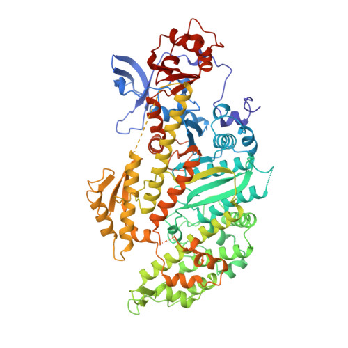
The three-dimensional structures of the truncated myosin head from Dictyostelium discoideum myosin II (S1dC) complexed with MgAMPPNP, MgATPgammaS, and MgADP are reported at 2.1, 1.9, and 2.1 A resolution, respectively. Crystals were obtained by cocrystallization and were isomorphous with respect to those of S1dC. MgADP.BeFx [Fisher, A. J., et al. (1995) Biochemistry 34, 8960-8972]. In all three structures, the electron density for the entire nucleotide was clearly discernible. The overall structures of all three complexes are very similar to that of the beryllium fluoride complex which suggests that the differences in the physiological effects of ATPgammaS and AMPPNP are due to the changes in the equilibrium between the actin-bound and actin-free states of myosin caused by the lower affinity of AMPPNP for myosin. In S1dC.MgAMPPNP, the presence of the bridging nitrogen prompts the side chain of Asn233 to rotate which disrupts the hydrogen bonding pattern in the nucleotide binding pocket and alters the water structure surrounding the ribose hydroxyl groups. It appears that this change is responsible for the reduced affinity of AMPPNP for myosin relative to ATPgammaS. In contrast to the G-proteins, there is no major change in the conformation of the ligands that coordinate the nucleotide in S1dC.MgADP. This is due to three water molecules that adopt the approximate positions of the three oxygens on the gamma-phosphate and maintain the interactions with the Mg2+ ion and protein molecule. Interestingly, the thiophosphate group is evident in S1dC.MgATPgammaS even though it is slowly hydrolyzed by myosin. This suggests that the conformation observed here and in chicken skeletal myosin subfragment-1 [Rayment, I., et al. (1993) Science 261, 50-58] is unable to hydrolyze ATP and represents the structure of the prehydrolysis weak binding state of myosin.
- Institute for Enzyme Research and Department of Biochemistry, University of Wisconsin, Madison, Wisconsin 53705, USA.
Organizational Affiliation: