1MLW | pdb_00001mlw

Crystal structure of human tryptophan hydroxylase with bound 7,8-dihydro-L-biopterin cofactor and Fe(III)


Experimental Data Snapshot

  • Method: X-RAY DIFFRACTION
  • Resolution: 1.71 Å
  • R-Value Free: 
    0.230 (Depositor), 0.229 (DCC) 
  • R-Value Work: 
    0.205 (Depositor), 0.206 (DCC) 

Starting Model: experimental
View more details

wwPDB Validation 3D Report Full Report

Validation slider image for 1MLW

Ligand Structure Quality Assessment 


This is version 1.3 of the entry. See complete history

Literature

Three-dimensional structure of human tryptophan hydroxylase and its implications for the biosynthesis of the neurotransmitters serotonin and melatonin

Wang, L.Erlandsen, H.Haavik, J.Knappskog, P.M.Stevens, R.C.

(2002) Biochemistry 41: 12569-12574

  • DOI: https://doi.org/10.1021/bi026561f
  • Primary Citation Related Structures: 
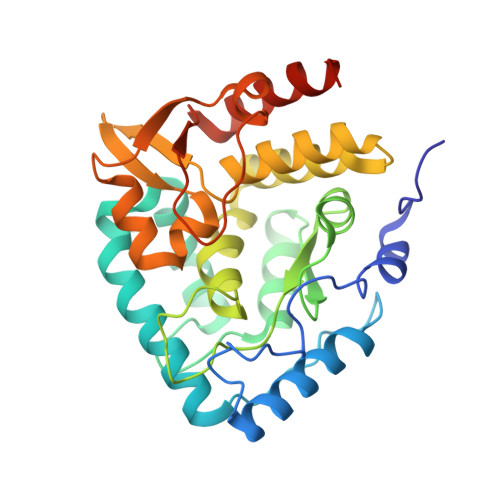
    1MLW

  • PubMed Abstract: 

    Tryptophan hydroxylase oxidizes L-tryptophan to 5-hydroxy-L-tryptophan in the rate-determining step of serotonin biosynthesis. We have determined the X-ray crystal structure (1.7 A) of a truncated functional form of human tryptophan hydroxylase with the bound cofactor analogue 7,8-dihydro-L-biopterin, providing the first atomic-resolution information for the catalytic domain of this important enzyme. Comparison of the three-dimensional structures of all three members of the aromatic amino acid hydroxylase family--tyrosine hydroxylase, phenylalanine hydroxylase, and tryptophan hydroxylase--reveals important differences at the active sites.


  • Organizational Affiliation
    • Department of Molecular Biology, The Scripps Research Institute, 10550 North Torrey Pines Road, La Jolla, California 92037, USA.

Macromolecule Content 

  • Total Structure Weight: 34.94 kDa 
  • Atom Count: 2,605 
  • Modeled Residue Count: 290 
  • Deposited Residue Count: 301 
  • Unique protein chains: 1

Macromolecules

Find similar proteins by:|  3D Structure
Entity ID: 1
MoleculeChains  Sequence LengthOrganismDetailsImage
Tryptophan 5-monooxygenase301Homo sapiensMutation(s): 0 
Gene Names: TPH
EC: 1.14.16.4
UniProt & NIH Common Fund Data Resources
Find proteins for P17752 (Homo sapiens)
Explore P17752 
Go to UniProtKB:  P17752
PHAROS:  P17752
GTEx:  ENSG00000129167 
Entity Groups
Sequence Clusters30% Identity50% Identity70% Identity90% Identity95% Identity100% Identity
UniProt GroupP17752
Sequence Annotations
Expand
Reference Sequence

Experimental Data & Validation

Experimental Data

  • Method: X-RAY DIFFRACTION
  • Resolution: 1.71 Å
  • R-Value Free:  0.230 (Depositor), 0.229 (DCC) 
  • R-Value Work:  0.205 (Depositor), 0.206 (DCC) 
Space Group: P 21 21 21
Unit Cell:
Length ( Å )Angle ( ˚ )
a = 47.775α = 90
b = 57.334β = 90
c = 109.41γ = 90
Software Package:
Software NamePurpose
HKL-2000data collection
SCALEPACKdata scaling
EPMRphasing
CNSrefinement
HKL-2000data reduction

Structure Validation

View Full Validation Report


Ligand Structure Quality Assessment 


Entry History 

Deposition Data

Revision History  (Full details and data files)

  • Version 1.0: 2002-12-18
    Type: Initial release
  • Version 1.1: 2007-10-16
    Changes: Version format compliance
  • Version 1.2: 2011-07-13
    Changes: Version format compliance
  • Version 1.3: 2024-02-14
    Changes: Data collection, Database references, Derived calculations, Refinement description