1MIO | pdb_00001mio

X-RAY CRYSTAL STRUCTURE OF THE NITROGENASE MOLYBDENUM-IRON PROTEIN FROM CLOSTRIDIUM PASTEURIANUM AT 3.0 ANGSTROMS RESOLUTION


Experimental Data Snapshot

  • Method: X-RAY DIFFRACTION
  • Resolution: 3.00 Å
  • R-Value Work: 
    0.180 (Depositor) 
  • R-Value Observed: 
    0.180 (Depositor) 

wwPDB Validation 3D Report Full Report

Validation slider image for 1MIO

This is version 1.3 of the entry. See complete history

Literature

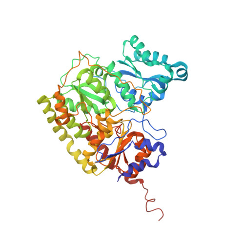
X-ray crystal structure of the nitrogenase molybdenum-iron protein from Clostridium pasteurianum at 3.0-A resolution.

Kim, J.Woo, D.Rees, D.C.

(1993) Biochemistry 32: 7104-7115

  • DOI: https://doi.org/10.1021/bi00079a006
  • Primary Citation Related Structures: 
    1MIO

  • PubMed Abstract: 

    The crystal structure of the nitrogenase molybdenum-iron (MoFe) protein from Clostridium pasteurianum (Cp1) has been determined at 3.0-A resolution by a combination of isomorphous replacement, molecular replacement, and noncrystallographic symmetry averaging. The structure of Cp1, including the two types of metal centers associated with the protein (the FeMo-cofactor and the P-cluster pair), is similar to that previously described for the MoFe-protein from Azotobacter vinelandii (Av1). Unique features of the Cp1 structure arise from the presence of an approximately 50-residue insertion in the alpha subunit and an approximately 50-residue deletion in the beta subunit. As a consequence, the FeMo-cofactor is more buried in Cp1 than in Av1, since the insertion is located on the surface above the FeMo-cofactor. The location of this insertion near the putative nitrogenase iron protein binding site provides a structural basis for the observation that the nitrogenase proteins from C. pasteurianum have low activity with complementary nitrogenase proteins isolated from other organisms. Mechanistic implications of the Cp1 structure for substrate entry/product release, substrate binding to the FeMo-cofactor, and electron- and proton-transfer reactions of nitrogenase are discussed.


  • Organizational Affiliation
    • Division of Chemistry and Chemical Engineering, California Institute of Technology, Pasadena 91125.

Macromolecule Content 

  • Total Structure Weight: 221.94 kDa 
  • Atom Count: 15,269 
  • Modeled Residue Count: 1,964 
  • Deposited Residue Count: 1,982 
  • Unique protein chains: 2

Macromolecules

Find similar proteins by:|  3D Structure
Entity ID: 1
MoleculeChains  Sequence LengthOrganismDetailsImage
NITROGENASE MOLYBDENUM IRON PROTEIN (ALPHA CHAIN)
A, C
533Clostridium pasteurianumMutation(s): 0 
EC: 1.18.6.1
UniProt
Find proteins for P00467 (Clostridium pasteurianum)
Explore P00467 
Go to UniProtKB:  P00467
Entity Groups
Sequence Clusters30% Identity50% Identity70% Identity90% Identity95% Identity100% Identity
UniProt GroupP00467
Sequence Annotations
Expand
Reference Sequence
Find similar proteins by:|  3D Structure
Entity ID: 2
MoleculeChains  Sequence LengthOrganismDetailsImage
NITROGENASE MOLYBDENUM IRON PROTEIN (BETA CHAIN)
B, D
458Clostridium pasteurianumMutation(s): 0 
EC: 1.18.6.1
UniProt
Find proteins for P11347 (Clostridium pasteurianum)
Explore P11347 
Go to UniProtKB:  P11347
Entity Groups
Sequence Clusters30% Identity50% Identity70% Identity90% Identity95% Identity100% Identity
UniProt GroupP11347
Sequence Annotations
Expand
Reference Sequence

Small Molecules

Ligands 4 Unique
IDChains Name / Formula / InChI Key2D Diagram3D Interactions
CFM

Query on CFM


Download:Ideal Coordinates CCD File
G [auth B],
K [auth D]
FE-MO-S CLUSTER
Fe7 Mo S9
UZRXIPMKRKMLQF-UHFFFAOYSA-N
CLP

Query on CLP


Download:Ideal Coordinates CCD File
H [auth B],
L [auth D]
FE-S CLUSTER
Fe8 S8
MJXAKWFPOPRKKV-UHFFFAOYSA-N
HCA

Query on HCA


Download:Ideal Coordinates CCD File
F [auth B],
J [auth D]
3-HYDROXY-3-CARBOXY-ADIPIC ACID
C7 H10 O7
XKJVEVRQMLKSMO-SSDOTTSWSA-N
CA

Query on CA


Download:Ideal Coordinates CCD File
E [auth B],
I [auth D]
CALCIUM ION
Ca
BHPQYMZQTOCNFJ-UHFFFAOYSA-N

Experimental Data & Validation

Experimental Data

  • Method: X-RAY DIFFRACTION
  • Resolution: 3.00 Å
  • R-Value Work:  0.180 (Depositor) 
  • R-Value Observed: 0.180 (Depositor) 
Space Group: P 1 21 1
Unit Cell:
Length ( Å )Angle ( ˚ )
a = 69.96α = 90
b = 151.3β = 110.4
c = 121.9γ = 90
Software Package:
Software NamePurpose
X-PLORmodel building
X-PLORrefinement
X-PLORphasing

Structure Validation

View Full Validation Report


Entry History 

Deposition Data

Revision History  (Full details and data files)

  • Version 1.0: 1993-10-31
    Type: Initial release
  • Version 1.1: 2008-03-24
    Changes: Version format compliance
  • Version 1.2: 2011-07-13
    Changes: Version format compliance
  • Version 1.3: 2024-02-14
    Changes: Data collection, Database references, Derived calculations, Other