1MHD

CRYSTAL STRUCTURE OF A SMAD MH1 DOMAIN BOUND TO DNA


Experimental Data Snapshot

  • Method: X-RAY DIFFRACTION
  • Resolution: 2.80 Å
  • R-Value Free: 0.288 
  • R-Value Work: 0.212 
  • R-Value Observed: 0.212 

wwPDB Validation   3D Report Full Report


This is version 1.2 of the entry. See complete history


Literature

Crystal structure of a Smad MH1 domain bound to DNA: insights on DNA binding in TGF-beta signaling.

Shi, Y.Wang, Y.F.Jayaraman, L.Yang, H.Massague, J.Pavletich, N.P.

(1998) Cell 94: 585-594

  • DOI: https://doi.org/10.1016/s0092-8674(00)81600-1
  • Primary Citation of Related Structures:  
    1MHD

  • PubMed Abstract: 

    The Smad family of proteins, which are frequently targeted by tumorigenic mutations in cancer, mediate TGF-beta signaling from cell membrane to nucleus. The crystal structure of a Smad3 MH1 domain bound to an optimal DNA sequence determined at 2.8 A resolution reveals a novel DNA-binding motif. In the crystals, base-specific DNA recognition is provided exclusively by a conserved 11-residue beta hairpin that is embedded in the major groove of DNA. A surface loop region, to which tumorigenic mutations map, has been identified as a functional surface important for Smad activity. This structure establishes a framework for understanding how Smad proteins may act in concert with other transcription factors in the regulation of TGF-beta-responsive genes.


  • Organizational Affiliation

    Department of Molecular Biology, Princeton University, Lewis Thomas Laboratory, New Jersey 08544, USA. ygshi@princeton.edu


Macromolecules

Find similar proteins by:  (by identity cutoff)  |  3D Structure
Entity ID: 3
MoleculeChains Sequence LengthOrganismDetailsImage
SMAD3C [auth A],
D [auth B]
132Homo sapiensMutation(s): 0 
Gene Names: SMAD
UniProt & NIH Common Fund Data Resources
Find proteins for P84022 (Homo sapiens)
Explore P84022 
Go to UniProtKB:  P84022
PHAROS:  P84022
GTEx:  ENSG00000166949 
Entity Groups  
Sequence Clusters30% Identity50% Identity70% Identity90% Identity95% Identity100% Identity
UniProt GroupP84022
Sequence Annotations
Expand
  • Reference Sequence

Find similar nucleic acids by:  Sequence   |   3D Structure  

Entity ID: 1
MoleculeChains LengthOrganismImage
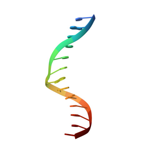
DNAA [auth C]13N/A
Sequence Annotations
Expand
  • Reference Sequence

Find similar nucleic acids by:  Sequence   |   3D Structure  

Entity ID: 2
MoleculeChains LengthOrganismImage
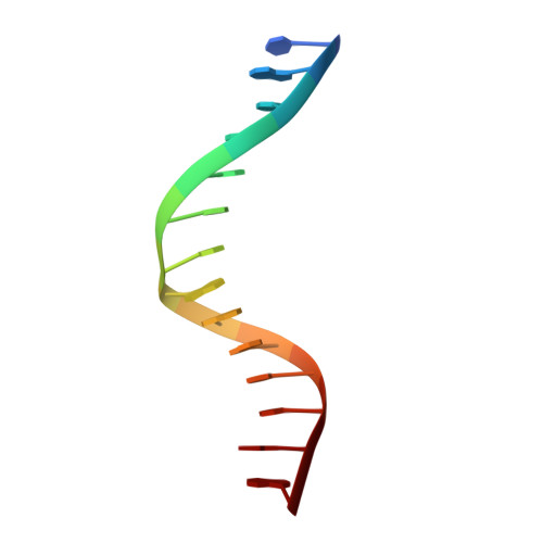
DNAB [auth D]14N/A
Sequence Annotations
Expand
  • Reference Sequence
Small Molecules
Binding Affinity Annotations 
IDSourceBinding Affinity
DNA PDBBind:  1MHD Kd: 114 (nM) from 1 assay(s)
Experimental Data & Validation

Experimental Data

  • Method: X-RAY DIFFRACTION
  • Resolution: 2.80 Å
  • R-Value Free: 0.288 
  • R-Value Work: 0.212 
  • R-Value Observed: 0.212 
  • Space Group: P 1 21 1
Unit Cell:
Length ( Å )Angle ( ˚ )
a = 45.6α = 90
b = 60.4β = 102
c = 71.6γ = 90
Software Package:
Software NamePurpose
DENZOdata reduction
SCALEPACKdata scaling
X-PLORmodel building
X-PLORrefinement
X-PLORphasing

Structure Validation

View Full Validation Report


Entry History 

Deposition Data

  • Released Date: 1999-08-18 
  • Deposition Author(s): Shi, Y.

Revision History  (Full details and data files)

  • Version 1.0: 1999-08-18
    Type: Initial release
  • Version 1.1: 2008-05-22
    Changes: Version format compliance
  • Version 1.2: 2011-07-13
    Changes: Version format compliance