1ME5 | pdb_00001me5

Crystal Structure of Mycobacterium Tuberculosis Alkylperoxidase AhpD H132Q Mutant


Experimental Data Snapshot

  • Method: X-RAY DIFFRACTION
  • Resolution: 2.40 Å
  • R-Value Free: 
    0.350 (Depositor) 
  • R-Value Work: 
    0.223 (Depositor) 
  • R-Value Observed: 
    0.235 (Depositor) 

Starting Model: experimental
View more details

wwPDB Validation 3D Report Full Report

Validation slider image for 1ME5

This is version 1.5 of the entry. See complete history

Literature

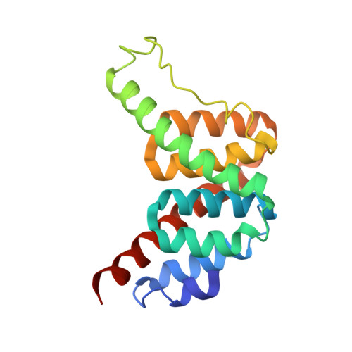
The mechanism of Mycobacterium tuberculosis alkylhydroperoxidase AhpD as defined by mutagenesis, crystallography, and kinetics.

Koshkin, A.Nunn, C.M.Djordjevic, S.Ortiz de Montellano, P.R.

(2003) J Biological Chem 278: 29502-29508

  • DOI: https://doi.org/10.1074/jbc.M303747200
  • Primary Citation Related Structures: 
    1LW1, 1ME5

  • PubMed Abstract: 

    AhpD, a protein with two cysteine residues, is required for physiological reduction of the Mycobacterium tuberculosis alkylhydroperoxidase AhpC. AhpD also has an alkylhydroperoxidase activity of its own. The AhpC/AhpD system provides critical antioxidant protection, particularly in the absence of the catalase-peroxidase KatG, which is suppressed in most isoniazid-resistant strains. Based on the crystal structure, we proposed recently a catalytic mechanism for AhpD involving a proton relay in which the Glu118 carboxylate group, via His137 and a water molecule, deprotonates the catalytic residue Cys133 (Nunn, C. M., Djordjevic, S., Hillas, P. J., Nishida, C., and Ortiz de Montellano, P. R. (2002) J. Biol. Chem. 277, 20033-20040). A possible role for His132 in subsequent formation of the Cys133-Cys130 disulfide bond was also noted. To test this proposed mechanism, we have expressed the H137F, H137Q, H132F, H132Q, E118F, E118Q, C133S, and C130S mutants of AhpD, determined the crystal structures of the H137F and H132Q mutants, estimated the pKa values of the cysteine residues, and defined the kinetic properties of the mutant proteins. The collective results strongly support the proposed catalytic mechanism for AhpD.


  • Organizational Affiliation
    • Department of Pharmaceutical Chemistry, University of California, San Francisco, California 94143-2280, USA.

Macromolecule Content 

  • Total Structure Weight: 56.37 kDa 
  • Atom Count: 4,215 
  • Modeled Residue Count: 515 
  • Deposited Residue Count: 531 
  • Unique protein chains: 1

Macromolecules

Find similar proteins by:|  3D Structure
Entity ID: 1
MoleculeChains  Sequence LengthOrganismDetailsImage
ALKYLHYDROPEROXIDASE D
A, B, C
177Mycobacterium tuberculosisMutation(s): 1 
EC: 1.11.1.28
UniProt
Find proteins for P9WQB5 (Mycobacterium tuberculosis (strain ATCC 25618 / H37Rv))
Explore P9WQB5 
Go to UniProtKB:  P9WQB5
Entity Groups
Sequence Clusters30% Identity50% Identity70% Identity90% Identity95% Identity100% Identity
UniProt GroupP9WQB5
Sequence Annotations
Expand
Reference Sequence

Experimental Data & Validation

Experimental Data

  • Method: X-RAY DIFFRACTION
  • Resolution: 2.40 Å
  • R-Value Free:  0.350 (Depositor) 
  • R-Value Work:  0.223 (Depositor) 
  • R-Value Observed: 0.235 (Depositor) 
Space Group: C 1 2 1
Unit Cell:
Length ( Å )Angle ( ˚ )
a = 100.119α = 90
b = 58.652β = 120.47
c = 88.914γ = 90
Software Package:
Software NamePurpose
DENZOdata reduction
SCALEPACKdata scaling
CNSrefinement
CNSphasing

Structure Validation

View Full Validation Report


Entry History 

Deposition Data

Revision History  (Full details and data files)

  • Version 1.0: 2002-09-11
    Type: Initial release
  • Version 1.1: 2008-04-28
    Changes: Version format compliance
  • Version 1.2: 2011-07-13
    Changes: Version format compliance
  • Version 1.3: 2018-01-31
    Changes: Advisory, Experimental preparation
  • Version 1.4: 2021-10-27
    Changes: Advisory, Database references
  • Version 1.5: 2024-02-14
    Changes: Data collection, Refinement description