1M5N | pdb_00001m5n

Crystal structure of HEAT repeats (1-11) of importin b bound to the non-classical NLS(67-94) of PTHrP


Experimental Data Snapshot

  • Method: X-RAY DIFFRACTION
  • Resolution: 2.90 Å
  • R-Value Free: 
    0.297 (Depositor) 
  • R-Value Work: 
    0.250 (Depositor) 
  • R-Value Observed: 
    0.250 (Depositor) 

Starting Model: experimental
View more details

wwPDB Validation 3D Report Full Report

Validation slider image for 1M5N

This is version 1.3 of the entry. See complete history

Literature

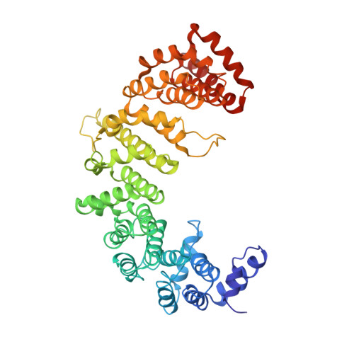
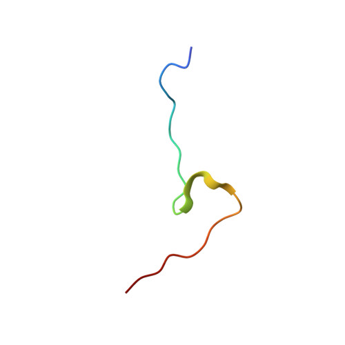
Molecular basis for the recognition of a nonclassical nuclear localization signal by importin beta

Cingolani, G.Bednenko, J.Gillespie, M.T.Gerace, L.

(2002) Mol Cell 10: 1345-1353

  • DOI: https://doi.org/10.1016/s1097-2765(02)00727-x
  • Primary Citation Related Structures: 
    1M5N

  • PubMed Abstract: 

    Nuclear import of proteins containing a classical nuclear localization signal (NLS) involves NLS recognition by importin alpha, which associates with importin beta via the IBB domain. Other proteins, including parathyroid hormone-related protein (PTHrP), are imported into the nucleus by direct interaction with importin beta. We solved the crystal structure of a fragment of importin beta-1 (1-485) bound to the nonclassical NLS of PTHrP. The structure reveals a second extended cargo binding site on importin beta distinct from the IBB domain binding site. Using a permeabilized cell import assay we demonstrate that importin beta (1-485) can import PTHrP-coupled cargo in a Ran-dependent manner. We propose that this region contains a prototypical nuclear import receptor domain, which could have evolved into the modern importin beta superfamily.


  • Organizational Affiliation
    • Departments of Cell and Molecular Biology, The Scripps Research Institute, La Jolla, CA 92037, USA.

Macromolecule Content 

  • Total Structure Weight: 57.17 kDa 
  • Atom Count: 4,020 
  • Modeled Residue Count: 513 
  • Deposited Residue Count: 513 
  • Unique protein chains: 2

Macromolecules

Find similar proteins by:|  3D Structure
Entity ID: 1
MoleculeChains  Sequence LengthOrganismDetailsImage
Importin beta-1 subunitA [auth S]485Homo sapiensMutation(s): 0 
UniProt & NIH Common Fund Data Resources
Find proteins for Q14974 (Homo sapiens)
Explore Q14974 
Go to UniProtKB:  Q14974
PHAROS:  Q14974
GTEx:  ENSG00000108424 
Entity Groups
Sequence Clusters30% Identity50% Identity70% Identity90% Identity95% Identity100% Identity
UniProt GroupQ14974
Sequence Annotations
Expand
Reference Sequence
Find similar proteins by:|  3D Structure
Entity ID: 2
MoleculeChains  Sequence LengthOrganismDetailsImage
Parathyroid hormone-related proteinB [auth Q]28N/AMutation(s): 0 
UniProt & NIH Common Fund Data Resources
Find proteins for P12272 (Homo sapiens)
Explore P12272 
Go to UniProtKB:  P12272
PHAROS:  P12272
GTEx:  ENSG00000087494 
Entity Groups
Sequence Clusters30% Identity50% Identity70% Identity90% Identity95% Identity100% Identity
UniProt GroupP12272
Sequence Annotations
Expand
Reference Sequence

Experimental Data & Validation

Experimental Data

  • Method: X-RAY DIFFRACTION
  • Resolution: 2.90 Å
  • R-Value Free:  0.297 (Depositor) 
  • R-Value Work:  0.250 (Depositor) 
  • R-Value Observed: 0.250 (Depositor) 
Space Group: C 2 2 21
Unit Cell:
Length ( Å )Angle ( ˚ )
a = 92.792α = 90
b = 105.447β = 90
c = 146.738γ = 90
Software Package:
Software NamePurpose
DENZOdata reduction
SCALEPACKdata scaling
AMoREphasing
CNSrefinement

Structure Validation

View Full Validation Report


Entry History 

Deposition Data

Revision History  (Full details and data files)

  • Version 1.0: 2003-01-21
    Type: Initial release
  • Version 1.1: 2008-04-28
    Changes: Version format compliance
  • Version 1.2: 2011-07-13
    Changes: Version format compliance
  • Version 1.3: 2024-02-14
    Changes: Data collection, Database references, Refinement description