1M4G

Aminoglycoside 2'-N-acetyltransferase from Mycobacterium tuberculosis-Complex with Coenzyme A and Ribostamycin


Experimental Data Snapshot

  • Method: X-RAY DIFFRACTION
  • Resolution: 1.80 Å
  • R-Value Free: 0.202 
  • R-Value Work: 0.165 

wwPDB Validation   3D Report Full Report


Ligand Structure Quality Assessment 


This is version 1.3 of the entry. See complete history


Literature

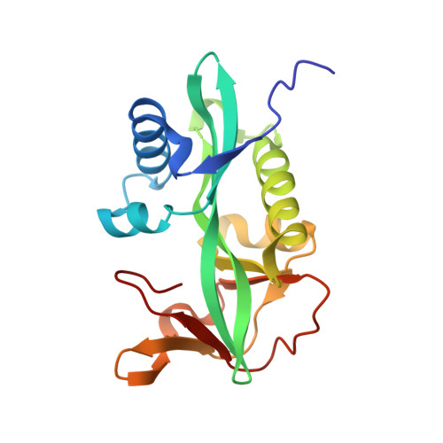
Aminoglycoside 2'-N-acetyltransferase from Mycobacterium tuberculosis in complex with coenzyme A and aminoglycoside substrates.

Vetting, M.W.Hegde, S.S.Javid-Majd, F.Blanchard, J.S.Roderick, S.L.

(2002) Nat Struct Biol 9: 653-658

  • DOI: https://doi.org/10.1038/nsb830
  • Primary Citation of Related Structures:  
    1M44, 1M4D, 1M4G, 1M4I

  • PubMed Abstract: 

    AAC(2')-Ic catalyzes the coenzyme A (CoA)-dependent acetylation of the 2' hydroxyl or amino group of a broad spectrum of aminoglycosides. The crystal structure of the AAC(2')-Ic from Mycobacterium tuberculosis has been determined in the apo enzyme form and in ternary complexes with CoA and either tobramycin, kanamycin A or ribostamycin, representing the first structures of an aminoglycoside acetyltransferase bound to a drug. The overall fold of AAC(2')-Ic places it in the GCN5-related N-acetyltransferase (GNAT) superfamily. Although the physiological function of AAC(2')-Ic is uncertain, a structural analysis of these high-affinity aminoglycoside complexes suggests that the enzyme may acetylate a key biosynthetic intermediate of mycothiol, the major reducing agent in mycobacteria, and participate in the regulation of cellular redox potential.


  • Organizational Affiliation

    Department of Biochemistry, Albert Einstein College of Medicine, 1300 Morris Park Avenue, Bronx, New York 10461, USA.


Macromolecules
Find similar proteins by:  (by identity cutoff)  |  3D Structure
Entity ID: 1
MoleculeChains Sequence LengthOrganismDetailsImage
Aminoglycoside 2'-N-acetyltransferase
A, B
181Mycobacterium tuberculosis H37RvMutation(s): 0 
EC: 2.3.1
UniProt
Find proteins for P9WQG9 (Mycobacterium tuberculosis (strain ATCC 25618 / H37Rv))
Explore P9WQG9 
Go to UniProtKB:  P9WQG9
Entity Groups  
Sequence Clusters30% Identity50% Identity70% Identity90% Identity95% Identity100% Identity
UniProt GroupP9WQG9
Sequence Annotations
Expand
  • Reference Sequence
Experimental Data & Validation

Experimental Data

  • Method: X-RAY DIFFRACTION
  • Resolution: 1.80 Å
  • R-Value Free: 0.202 
  • R-Value Work: 0.165 
  • Space Group: P 21 21 21
Unit Cell:
Length ( Å )Angle ( ˚ )
a = 48.7α = 90
b = 86.4β = 90
c = 98.4γ = 90
Software Package:
Software NamePurpose
AMoREphasing
CNSrefinement
DENZOdata reduction
SCALEPACKdata scaling

Structure Validation

View Full Validation Report


Ligand Structure Quality Assessment 


Entry History 

Deposition Data

Revision History  (Full details and data files)

  • Version 1.0: 2002-08-28
    Type: Initial release
  • Version 1.1: 2008-04-28
    Changes: Version format compliance
  • Version 1.2: 2011-07-13
    Changes: Source and taxonomy, Version format compliance
  • Version 1.3: 2024-02-14
    Changes: Data collection, Database references, Derived calculations, Refinement description, Structure summary