1M40 | pdb_00001m40

ULTRA HIGH RESOLUTION CRYSTAL STRUCTURE OF TEM-1


Experimental Data Snapshot

  • Method: X-RAY DIFFRACTION
  • Resolution: 0.85 Å
  • R-Value Free: 
    0.112 (Depositor), 0.110 (DCC) 
  • R-Value Work: 
    0.091 (Depositor), 0.092 (DCC) 

Starting Model: experimental
View more details

wwPDB Validation 3D Report Full Report

Validation slider image for 1M40

Ligand Structure Quality Assessment 


This is version 1.4 of the entry. See complete history

Literature

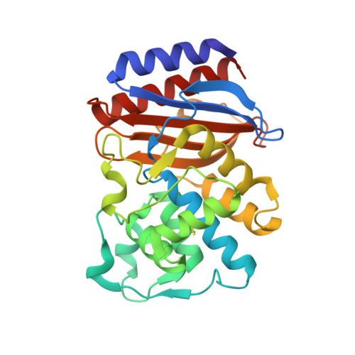
An ultrahigh resolution structure of TEM-1 beta-lactamase suggests a role for Glu166 as the general base in acylation.

Minasov, G.Wang, X.Shoichet, B.K.

(2002) J Am Chem Soc 124: 5333-5340

  • DOI: https://doi.org/10.1021/ja0259640
  • Primary Citation Related Structures: 
    1M40

  • PubMed Abstract: 

    Although TEM-1 beta-lactamase is among the best studied enzymes, its acylation mechanism remains controversial. To investigate this problem, the structure of TEM-1 in complex with an acylation transition-state analogue was determined at ultrahigh resolution (0.85 A) by X-ray crystallography. The quality of the data was such as to allow for refinement to an R-factor of 9.1% and an R(free) of 11.2%. In the resulting structure, the electron density features were clear enough to differentiate between single and double bonds in carboxylate groups, to identify multiple conformations that are occupied by residues and loops, and to assign 70% of the protons in the protein. Unexpectedly, even at pH 8.0 where the protein was crystallized, the active site residue Glu166 is clearly protonated. This supports the hypothesis that Glu166 is the general base in the acylation half of the reaction cycle. This structure suggests that Glu166 acts through the catalytic water to activate Ser70 for nucleophilic attack on the beta-lactam ring of the substrate. The hydrolytic mechanism of class A beta-lactamases, such as TEM-1, appears to be symmetrical, as are the serine proteases. Apart from its mechanistic implications, this atomic resolution structure affords an unusually detailed view of the structure, dynamics, and hydrogen-bonding networks of TEM-1, which may be useful for the design of inhibitors against this key antibiotic resistance target.


  • Organizational Affiliation
    • Department of Molecular Pharmacology & Biological Chemistry, Northwestern University, 303 East Chicago Avenue, Chicago, Illinois 60611-3008, USA.

Macromolecule Content 

  • Total Structure Weight: 29.68 kDa 
  • Atom Count: 3,969 
  • Modeled Residue Count: 263 
  • Deposited Residue Count: 263 
  • Unique protein chains: 1

Macromolecules

Find similar proteins by:|  3D Structure
Entity ID: 1
MoleculeChains  Sequence LengthOrganismDetailsImage
BETA-LACTAMASE TEM263Escherichia coliMutation(s): 1 
Gene Names: bla
EC: 3.5.2.6
UniProt
Find proteins for P62593 (Escherichia coli)
Explore P62593 
Go to UniProtKB:  P62593
Entity Groups
Sequence Clusters30% Identity50% Identity70% Identity90% Identity95% Identity100% Identity
UniProt GroupP62593
Sequence Annotations
Expand
Reference Sequence

Small Molecules

Ligands 3 Unique
IDChains Name / Formula / InChI Key2D Diagram3D Interactions
CB4

Query on CB4


Download:Ideal Coordinates CCD File
I [auth A]PINACOL[[2-AMINO-ALPHA-(1-CARBOXY-1-METHYLETHOXYIMINO)-4-THIAZOLEACETYL]AMINO]METHANEBORONATE
C10 H15 B N4 O6 S
ZECCQELUYUPTSB-UUASQNMZSA-N
PO4

Query on PO4


Download:Ideal Coordinates CCD File
B [auth A],
C [auth A],
D [auth A]
PHOSPHATE ION
O4 P
NBIIXXVUZAFLBC-UHFFFAOYSA-K
K

Query on K


Download:Ideal Coordinates CCD File
E [auth A],
F [auth A],
G [auth A],
H [auth A]
POTASSIUM ION
K
NPYPAHLBTDXSSS-UHFFFAOYSA-N

Experimental Data & Validation

Experimental Data

  • Method: X-RAY DIFFRACTION
  • Resolution: 0.85 Å
  • R-Value Free:  0.112 (Depositor), 0.110 (DCC) 
  • R-Value Work:  0.091 (Depositor), 0.092 (DCC) 
Space Group: P 21 21 21
Unit Cell:
Length ( Å )Angle ( ˚ )
a = 41.313α = 90
b = 61.641β = 90
c = 89.252γ = 90
Software Package:
Software NamePurpose
DENZOdata reduction
SCALEPACKdata scaling
AMoREphasing
SHELXL-97refinement

Structure Validation

View Full Validation Report


Ligand Structure Quality Assessment 


Entry History 

Deposition Data

Revision History  (Full details and data files)

  • Version 1.0: 2002-07-17
    Type: Initial release
  • Version 1.1: 2008-04-28
    Changes: Version format compliance
  • Version 1.2: 2011-07-13
    Changes: Version format compliance
  • Version 1.3: 2021-10-27
    Changes: Database references, Derived calculations
  • Version 1.4: 2024-11-20
    Changes: Data collection, Refinement description, Structure summary