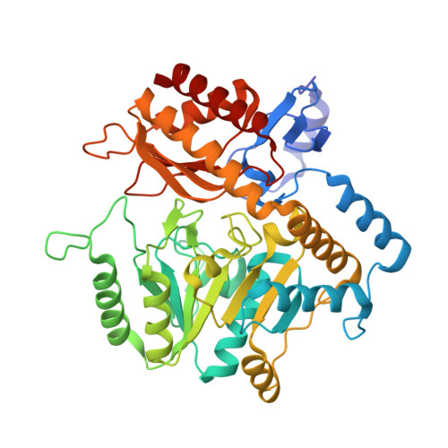
1M0P

Structure of Dialkylglycine Decarboxylase Complexed with 1-Amino-1-phenylethanephosphonate


Experimental Data Snapshot

  • Method: X-RAY DIFFRACTION
  • Resolution: 2.60 Å
  • R-Value Free: 0.230 
  • R-Value Work: 0.173 
  • R-Value Observed: 0.178 

Starting Model: experimental
View more details

wwPDB Validation   3D Report Full Report


This is version 1.4 of the entry. See complete history


Literature

Aminophosphonate Inhibitors of Dialkylglycine Decarboxylase: Structural Basis for Slow Binding Inhibition

Liu, W.Rogers, C.J.Fisher, A.J.Toney, M.D.

(2002) Biochemistry 41: 12320-12328

  • DOI: https://doi.org/10.1021/bi026318g
  • Primary Citation of Related Structures:  
    1M0N, 1M0O, 1M0P, 1M0Q

  • PubMed Abstract: 

    The kinetics of inhibition of dialkylglycine decarboxylase by five aminophosphonate inhibitors are presented. Two of these [(R)-1-amino-1-methylpropanephosphonate and (S)-1-aminoethanephosphonate] are slow binding inhibitors. The inhibitors follow a mechanism in which a weak complex is rapidly formed, followed by slow isomerization to the tight complex. Here, the tight complexes are bound 10-fold more tightly than the weak, initial complexes. The slow onset inhibition occurs with t(1/2) values of 1.3 and 0.55 min at saturating inhibitor concentrations for the AMPP and S-AEP inhibitors, respectively, while dissociation of these inhibitor complexes occurs with t(1/2) values of 13 and 4.6 min, respectively. The X-ray structures of four of the inhibitors in complex with dialkylglycine decarboxylase have been determined to resolutions ranging from 2.6 to 2.0 A, and refined to R-factors of 14.5-19.5%. These structures show variation in the active site structure with inhibitor side chain size and slow binding character. It is proposed that the slow binding behavior originates in an isomerization from an initial complex in which the PLP pyridine nitrogen-D243 OD2 distance is approximately 2.9 A to one in which it is approximately 2.7 A. The angles that the C-P bonds make with the p orbitals of the aldimine pi system are correlated with the reactivities of the analogous amino acid substrates, suggesting a role for stereoelectronic effects in Schiff base reactivity.


  • Organizational Affiliation

    Department of Chemistry, University of California, Davis, California 95616, USA.


Macromolecules
Find similar proteins by:  (by identity cutoff)  |  3D Structure
Entity ID: 1
MoleculeChains Sequence LengthOrganismDetailsImage
2,2-Dialkylglycine Decarboxylase433Burkholderia cepaciaMutation(s): 0 
EC: 4.1.1.64
UniProt
Find proteins for P16932 (Burkholderia cepacia)
Explore P16932 
Go to UniProtKB:  P16932
Entity Groups  
Sequence Clusters30% Identity50% Identity70% Identity90% Identity95% Identity100% Identity
UniProt GroupP16932
Sequence Annotations
Expand
  • Reference Sequence
Small Molecules
Ligands 3 Unique
IDChains Name / Formula / InChI Key2D Diagram3D Interactions
ELP
Query on ELP

Download Ideal Coordinates CCD File 
D [auth A](1R)-1-[((1E)-{3-HYDROXY-2-METHYL-5-[(PHOSPHONOOXY)METHYL]PYRIDIN-4-YL}METHYLENE)AMINO]-1-PHENYLETHYLPHOSPHONIC ACID
C16 H20 N2 O8 P2
YMGMBGLRDKSMMV-TWIVVOHQSA-N
K
Query on K

Download Ideal Coordinates CCD File 
B [auth A]POTASSIUM ION
K
NPYPAHLBTDXSSS-UHFFFAOYSA-N
NA
Query on NA

Download Ideal Coordinates CCD File 
C [auth A]SODIUM ION
Na
FKNQFGJONOIPTF-UHFFFAOYSA-N
Experimental Data & Validation

Experimental Data

  • Method: X-RAY DIFFRACTION
  • Resolution: 2.60 Å
  • R-Value Free: 0.230 
  • R-Value Work: 0.173 
  • R-Value Observed: 0.178 
  • Space Group: P 64 2 2
Unit Cell:
Length ( Å )Angle ( ˚ )
a = 150.77α = 90
b = 150.77β = 90
c = 84.63γ = 120
Software Package:
Software NamePurpose
TNTrefinement
SCALEPACKdata scaling
CNSrefinement
DENZOdata reduction
CNSphasing

Structure Validation

View Full Validation Report


Entry History 

Deposition Data

Revision History  (Full details and data files)

  • Version 1.0: 2002-10-23
    Type: Initial release
  • Version 1.1: 2008-04-28
    Changes: Version format compliance
  • Version 1.2: 2011-07-13
    Changes: Version format compliance
  • Version 1.3: 2019-07-24
    Changes: Data collection, Refinement description
  • Version 1.4: 2024-02-14
    Changes: Data collection, Database references, Derived calculations, Refinement description