1M0D

Crystal Structure of Bacteriophage T7 Endonuclease I with a Wild-Type Active Site and Bound Manganese Ions


Experimental Data Snapshot

  • Method: X-RAY DIFFRACTION
  • Resolution: 1.90 Å
  • R-Value Free: 0.234 
  • R-Value Work: 0.210 
  • R-Value Observed: 0.210 

Starting Model: experimental
View more details

wwPDB Validation   3D Report Full Report


This is version 1.3 of the entry. See complete history


Literature

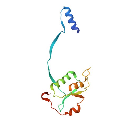
Metal ions bound at the active site of the junction-resolving enzyme T7 endonuclease I.

Hadden, J.M.Declais, A.C.Phillips, S.E.Lilley, D.M.

(2002) EMBO J 21: 3505-3515

  • DOI: https://doi.org/10.1093/emboj/cdf337
  • Primary Citation of Related Structures:  
    1M0D, 1M0I

  • PubMed Abstract: 

    T7 endonuclease I is a nuclease that is selective for the structure of the four-way DNA junction. The active site is similar to those of a number of restriction enzymes. We have solved the crystal structure of endonuclease I with a wild-type active site. Diffusion of manganese ions into the crystal revealed two peaks of electron density per active site, defining two metal ion-binding sites. Site 1 is fully occupied, and the manganese ion is coordinated by the carboxylate groups of Asp55 and Glu65, and the main chain carbonyl of Thr66. Site 2 is partially occupied, and the metal ion has a single protein ligand, the remaining carboxylate oxygen atom of Asp55. Isothermal titration calorimetry showed the sequential exothermic binding of two manganese ions in solution, with dissociation constants of 0.58 +/- 0.019 and 14 +/- 1.5 mM. These results are consistent with a two metal ion mechanism for the cleavage reaction, in which the hydrolytic water molecule is contained in the first coordination sphere of the site 1-bound metal ion.


  • Organizational Affiliation

    Astbury Centre for Structural Molecular Biology, School of Biochemistry and Molecular Biology, University of Leeds, Leeds LS2 9JT, UK.


Macromolecules
Find similar proteins by:  (by identity cutoff)  |  3D Structure
Entity ID: 1
MoleculeChains Sequence LengthOrganismDetailsImage
Endodeoxyribonuclease I
A, B, C, D
138Escherichia phage T7Mutation(s): 0 
Gene Names: Endonuclease I
EC: 3.1.21.2
UniProt
Find proteins for P00641 (Escherichia phage T7)
Explore P00641 
Go to UniProtKB:  P00641
Entity Groups  
Sequence Clusters30% Identity50% Identity70% Identity90% Identity95% Identity100% Identity
UniProt GroupP00641
Sequence Annotations
Expand
  • Reference Sequence
Small Molecules
Ligands 2 Unique
IDChains Name / Formula / InChI Key2D Diagram3D Interactions
SO4
Query on SO4

Download Ideal Coordinates CCD File 
G [auth A],
J [auth B],
M [auth C],
P [auth D]
SULFATE ION
O4 S
QAOWNCQODCNURD-UHFFFAOYSA-L
MN
Query on MN

Download Ideal Coordinates CCD File 
E [auth A]
F [auth A]
H [auth B]
I [auth B]
K [auth C]
E [auth A],
F [auth A],
H [auth B],
I [auth B],
K [auth C],
L [auth C],
N [auth D],
O [auth D]
MANGANESE (II) ION
Mn
WAEMQWOKJMHJLA-UHFFFAOYSA-N
Experimental Data & Validation

Experimental Data

  • Method: X-RAY DIFFRACTION
  • Resolution: 1.90 Å
  • R-Value Free: 0.234 
  • R-Value Work: 0.210 
  • R-Value Observed: 0.210 
  • Space Group: P 21 21 2
Unit Cell:
Length ( Å )Angle ( ˚ )
a = 123.81α = 90
b = 133.96β = 90
c = 61.46γ = 90
Software Package:
Software NamePurpose
CNSrefinement
MOSFLMdata reduction
CCP4data scaling
CNSphasing

Structure Validation

View Full Validation Report


Entry History 

Deposition Data

Revision History  (Full details and data files)

  • Version 1.0: 2002-07-10
    Type: Initial release
  • Version 1.1: 2008-04-28
    Changes: Version format compliance
  • Version 1.2: 2011-07-13
    Changes: Version format compliance
  • Version 1.3: 2024-02-14
    Changes: Data collection, Database references, Derived calculations, Refinement description