1LS2

Fitting of EF-Tu and tRNA in the Low Resolution Cryo-EM Map of an EF-Tu Ternary Complex (GDP and Kirromycin) Bound to E. coli 70S Ribosome


Experimental Data Snapshot

  • Method: ELECTRON MICROSCOPY
  • Resolution: 16.8 Å
  • Aggregation State: PARTICLE 
  • Reconstruction Method: SINGLE PARTICLE 

wwPDB Validation   3D Report Full Report


This is version 1.4 of the entry. See complete history


Literature

Cryo-EM reveals an active role for aminoacyl-tRNA in the accommodation process.

Valle, M.Sengupta, J.Swami, N.K.Grassucci, R.A.Burkhardt, N.Nierhaus, K.H.Agrawal, R.K.Frank, J.

(2002) EMBO J 21: 3557-3567

  • DOI: https://doi.org/10.1093/emboj/cdf326
  • Primary Citation of Related Structures:  
    1LS2, 1LU3

  • PubMed Abstract: 

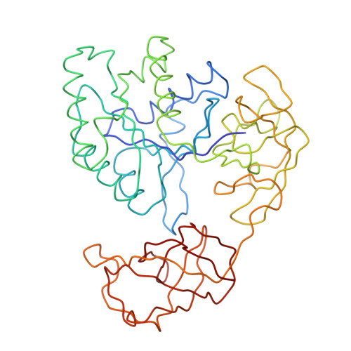
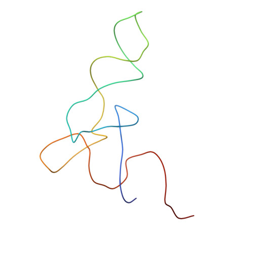
    During the elongation cycle of protein biosynthesis, the specific amino acid coded for by the mRNA is delivered by a complex that is comprised of the cognate aminoacyl-tRNA, elongation factor Tu and GTP. As this ternary complex binds to the ribosome, the anticodon end of the tRNA reaches the decoding center in the 30S subunit. Here we present the cryo- electron microscopy (EM) study of an Escherichia coli 70S ribosome-bound ternary complex stalled with an antibiotic, kirromycin. In the cryo-EM map the anticodon arm of the tRNA presents a new conformation that appears to facilitate the initial codon-anticodon interaction. Furthermore, the elbow region of the tRNA is seen to contact the GTPase-associated center on the 50S subunit of the ribosome, suggesting an active role of the tRNA in the transmission of the signal prompting the GTP hydrolysis upon codon recognition.


  • Organizational Affiliation

    Howard Hughes Medical Institute, Health Research, Inc., Empire State Plaza, Albany, NY 12201-0509, USA.


Macromolecules

Find similar proteins by:  (by identity cutoff)  |  3D Structure
Entity ID: 2
MoleculeChains Sequence LengthOrganismDetailsImage
Elongation Factor TuB [auth A]393Escherichia coliMutation(s): 0 
UniProt
Find proteins for P0CE48 (Escherichia coli (strain K12))
Explore P0CE48 
Go to UniProtKB:  P0CE48
Entity Groups  
Sequence Clusters30% Identity50% Identity70% Identity90% Identity95% Identity100% Identity
UniProt GroupP0CE48
Sequence Annotations
Expand
  • Reference Sequence
Find similar nucleic acids by:  (by identity cutoff)  |  3D Structure
Entity ID: 1
MoleculeChains LengthOrganismImage
Phenylalanine transfer RNAA [auth B]76Saccharomyces cerevisiae
Sequence Annotations
Expand
  • Reference Sequence
Experimental Data & Validation

Experimental Data

  • Method: ELECTRON MICROSCOPY
  • Resolution: 16.8 Å
  • Aggregation State: PARTICLE 
  • Reconstruction Method: SINGLE PARTICLE 
EM Software:
TaskSoftware PackageVersion
RECONSTRUCTIONSPIDER

Structure Validation

View Full Validation Report


Entry History 

Deposition Data

Revision History  (Full details and data files)

  • Version 1.0: 2002-06-26
    Type: Initial release
  • Version 1.1: 2008-04-28
    Changes: Version format compliance
  • Version 1.2: 2011-07-13
    Changes: Version format compliance
  • Version 1.3: 2018-07-18
    Changes: Data collection
  • Version 1.4: 2024-02-14
    Changes: Data collection, Database references, Refinement description