1LLS

CRYSTAL STRUCTURE OF UNLIGANDED MALTOSE BINDING PROTEIN WITH XENON


Experimental Data Snapshot

  • Method: X-RAY DIFFRACTION
  • Resolution: 1.80 Å
  • R-Value Free: 0.226 
  • R-Value Work: 0.204 
  • R-Value Observed: 0.204 

wwPDB Validation   3D Report Full Report


This is version 1.3 of the entry. See complete history


Literature

DETECTION AND CHARACTERIZATION OF XENON-BINDING SITES IN PROTEINS BY 129XE NMR SPECTROSCOPY

Rubin, S.M.Lee, S.-Y.Ruiz, E.J.Pines, A.Wemmer, D.E.

(2002) J Mol Biol 322: 425-440

  • DOI: https://doi.org/10.1016/s0022-2836(02)00739-8
  • Primary Citation of Related Structures:  
    1LLS

  • PubMed Abstract: 

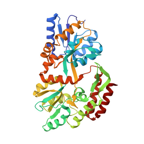
    Xenon-binding sites in proteins have led to a number of applications of xenon in biochemical and structural studies. Here we further develop the utility of 129Xe NMR in characterizing specific xenon-protein interactions. The sensitivity of the 129Xe chemical shift to its local environment and the intense signals attainable by optical pumping make xenon a useful NMR reporter of its own interactions with proteins. A method for detecting specific xenon-binding interactions by analysis of 129Xe chemical shift data is illustrated using the maltose binding protein (MBP) from Escherichia coli as an example. The crystal structure of MBP in the presence of 8atm of xenon confirms the binding site determined from NMR data. Changes in the structure of the xenon-binding cavity upon the binding of maltose by the protein can account for the sensitivity of the 129Xe chemical shift to MBP conformation. 129Xe NMR data for xenon in solution with a number of cavity containing phage T4 lysozyme mutants show that xenon can report on cavity structure. In particular, a correlation exists between cavity size and the binding-induced 129Xe chemical shift. Further applications of 129Xe NMR to biochemical assays, including the screening of proteins for xenon binding for crystallography are considered.


  • Organizational Affiliation

    Department of Chemistry, MC-1460, University of California, Berkeley 94720-1460, USA.


Macromolecules
Find similar proteins by:  (by identity cutoff)  |  3D Structure
Entity ID: 1
MoleculeChains Sequence LengthOrganismDetailsImage
Maltose-binding periplasmic protein370Escherichia coliMutation(s): 0 
UniProt
Find proteins for P0AEX9 (Escherichia coli (strain K12))
Explore P0AEX9 
Go to UniProtKB:  P0AEX9
Entity Groups  
Sequence Clusters30% Identity50% Identity70% Identity90% Identity95% Identity100% Identity
UniProt GroupP0AEX9
Sequence Annotations
Expand
  • Reference Sequence
Small Molecules
Ligands 1 Unique
IDChains Name / Formula / InChI Key2D Diagram3D Interactions
XE
Query on XE

Download Ideal Coordinates CCD File 
B [auth A]XENON
Xe
FHNFHKCVQCLJFQ-UHFFFAOYSA-N
Experimental Data & Validation

Experimental Data

  • Method: X-RAY DIFFRACTION
  • Resolution: 1.80 Å
  • R-Value Free: 0.226 
  • R-Value Work: 0.204 
  • R-Value Observed: 0.204 
  • Space Group: P 1
Unit Cell:
Length ( Å )Angle ( ˚ )
a = 38.207α = 100.75
b = 44.026β = 101.36
c = 57.649γ = 102.62
Software Package:
Software NamePurpose
CNSrefinement
MOSFLMdata reduction
CCP4data scaling
CNSphasing

Structure Validation

View Full Validation Report


Entry History 

Deposition Data

Revision History  (Full details and data files)

  • Version 1.0: 2002-09-18
    Type: Initial release
  • Version 1.1: 2008-04-28
    Changes: Version format compliance
  • Version 1.2: 2011-07-13
    Changes: Version format compliance
  • Version 1.3: 2023-08-16
    Changes: Data collection, Database references, Derived calculations, Refinement description