1LLF | pdb_00001llf

Cholesterol Esterase (Candida Cylindracea) Crystal Structure at 1.4A resolution


Experimental Data Snapshot

  • Method: X-RAY DIFFRACTION
  • Resolution: 1.40 Å
  • R-Value Free: 
    0.169 (Depositor), 0.170 (DCC) 
  • R-Value Work: 
    0.136 (Depositor), 0.140 (DCC) 
  • R-Value Observed: 
    0.142 (Depositor) 

Starting Model: experimental
View more details

wwPDB Validation   3D Report Full Report


Ligand Structure Quality Assessment 

Created with Raphaël 2.3.0Worse 01 BetterLigand structure goodness of fit to experimental dataBest fitted F23Click on this verticalbar to view details

This is version 2.2 of the entry. See complete history


Literature

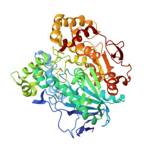
Three-dimensional structure of homodimeric cholesterol esterase-ligand complex at 1.4 A resolution.

Pletnev, V.Addlagatta, A.Wawrzak, Z.Duax, W.

(2003) Acta Crystallogr D Biol Crystallogr 59: 50-56

  • DOI: https://doi.org/10.1107/s0907444902018851
  • Primary Citation of Related Structures:  
    1LLF

  • PubMed Abstract: 

    The three-dimensional structure of a Candida cylindracea cholesterol esterase (ChE) homodimer (534 x 2 amino acids) in complex with a ligand of proposed formula C(23)H(48)O(2) has been determined at 1.4 A resolution in space group P1 using synchrotron low-temperature data. The structure refined to R = 0.136 and R(free) = 0.169 and has revealed new stereochemical details in addition to those detected for the apo- and holo-forms at 1.9 and 2.0 A resolution, respectively [Ghosh et al. (1995), Structure, 3, 279-288]. The cholesterol esterase structure is a dimer with four spatially separated interfacial contact areas and two symmetry-related pairs of openings to an internal intradimer cavity. Hydrophobic active-site gorges in each subunit face each other across a central interfacial cavity. The ChE subunits have carbohydrate chains attached to their Asn314 and Asn351 residues, with two ordered N-acetyl-D-glucosoamine moieties visible at each site. The side chains of 14 residues have two alternative conformations with occupancy values of 0.5 +/- 0.2. For each subunit the electron density in the enzyme active-site gorge is well modeled by a C(23)-chain fatty acid.


  • Organizational Affiliation

    Institute of Bioorganic Chemistry, Russian Academy of Science, Moscow, Russia.


Macromolecules
Find similar proteins by:  (by identity cutoff)  |  3D Structure
Entity ID: 1
MoleculeChains Sequence LengthOrganismDetailsImage
Lipase 3
A, B
534Limtongozyma cylindraceaMutation(s): 0 
EC: 3.1.1.3
UniProt
Find proteins for Q6S5M9 (Limtongozyma cylindracea)
Explore Q6S5M9 
Go to UniProtKB:  Q6S5M9
Entity Groups  
Sequence Clusters30% Identity50% Identity70% Identity90% Identity95% Identity100% Identity
UniProt GroupQ6S5M9
Glycosylation
Glycosylation Sites: 2
Sequence Annotations
Expand
  • Reference Sequence
Oligosaccharides

Help

Entity ID: 2
MoleculeChains Length2D Diagram Glycosylation3D Interactions
2-acetamido-2-deoxy-beta-D-glucopyranose-(1-4)-2-acetamido-2-deoxy-beta-D-glucopyranose
C, D, E, F
2N-Glycosylation
Glycosylation Resources
GlyTouCan:  G42666HT
GlyCosmos:  G42666HT
GlyGen:  G42666HT
Experimental Data & Validation

Experimental Data

  • Method: X-RAY DIFFRACTION
  • Resolution: 1.40 Å
  • R-Value Free:  0.169 (Depositor), 0.170 (DCC) 
  • R-Value Work:  0.136 (Depositor), 0.140 (DCC) 
  • R-Value Observed: 0.142 (Depositor) 
Space Group: P 1
Unit Cell:
Length ( Å )Angle ( ˚ )
a = 58.461α = 92.71
b = 58.477β = 97.48
c = 89.515γ = 109.38
Software Package:
Software NamePurpose
DENZOdata reduction
SCALEPACKdata scaling
AMoREphasing
SHELXL-97refinement

Structure Validation

View Full Validation Report


Ligand Structure Quality Assessment 

Created with Raphaël 2.3.0Worse 01 BetterLigand structure goodness of fit to experimental dataBest fitted F23Click on this verticalbar to view details

Entry History 

Deposition Data

Revision History  (Full details and data files)

  • Version 1.0: 2003-01-07
    Type: Initial release
  • Version 1.1: 2007-10-21
    Changes: Version format compliance
  • Version 1.2: 2011-07-13
    Changes: Non-polymer description, Version format compliance
  • Version 2.0: 2020-07-29
    Type: Remediation
    Reason: Carbohydrate remediation
    Changes: Advisory, Atomic model, Data collection, Derived calculations, Structure summary
  • Version 2.1: 2023-08-16
    Changes: Data collection, Database references, Refinement description, Structure summary
  • Version 2.2: 2024-11-13
    Changes: Structure summary