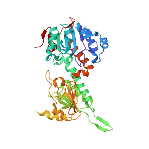
Crystal Structures of Transhydrogenase Domain I with and without Bound NADH
Prasad, G.S., Wahlberg, M., Sridhar, V., Yamaguchi, M., Hatefi, Y., Stout, C.D.(2002) Biochemistry 41: 12745-12754
- PubMed: 12379117 Search on PubMed
- DOI: https://doi.org/10.1021/bi020251f
- Primary Citation Related Structures:
1L7D, 1L7E - PubMed Abstract:
Transhydrogenase (TH) is a dimeric integral membrane enzyme in mitochondria and prokaryotes that couples proton translocation across a membrane with hydride transfer between NAD(H) and NADP(H) in soluble domains. Crystal structures of the NAD(H) binding alpha1 subunit (domain I) of Rhodospirillum rubrum TH have been determined at 1.8 A resolution in the absence of dinucleotide and at 1.9 A resolution with NADH bound. Each structure contains two domain I dimers in the asymmetric unit (AB and CD); the dimers are intimately associated and related by noncrystallographic 2-fold axes. NADH binds to subunits A and D, consistent with the half-of-the-sites reactivity of the enzyme. The conformation of NADH in subunits A and D is very similar; the nicotinamide is in the anti conformation, the A-face is exposed to solvent, and both N7 and O7 participate in hydrogen bonds. Comparison of subunits A and D to six independent copies of the subunit without bound NADH reveals multiple conformations for residues and loops surrounding the NADH site, indicating flexibility for binding and release of the substrate (product). The NADH-bound structure is also compared to the structures of R. rubrum domain I with NAD bound (PDB code 1F8G) and with NAD bound in complex with domain III of TH (PDB code 1HZZ). The NADH- vs NAD-bound domain I structures reveal conformational differences in conserved residues in the NAD(H) binding site and in dinucleotide conformation that are correlated with the net charge, i.e., oxidation state, of the nicotinamides. The comparisons illustrate how nicotinamide oxidation state can affect the domain I conformation, which is relevant to the hydride transfer step of the overall reaction.
- Division of Biochemistry, Department of Molecular and Experimental Medicine, The Scripps Research Institute, La Jolla, California 92037-1093, USA.
Organizational Affiliation: