Structure of Ala24/Asp61 --> Asp24/Asn61 substituted subunit c of Escherichia coli ATP synthase: implications for the mechanism of proton transport and rotary movement in the F0 complex.
Dmitriev, O.Y., Abildgaard, F., Markley, J.L., Fillingame, R.H.(2002) Biochemistry 41: 5537-5547
- PubMed: 11969414 Search on PubMed
- DOI: https://doi.org/10.1021/bi012198l
- Primary Citation Related Structures:
1L6T - PubMed Abstract:
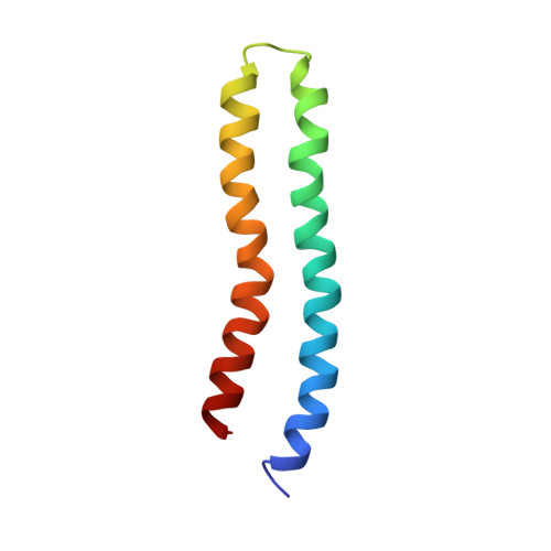
The structure of the A24D/D61N substituted subunit c of Escherichia coli ATP synthase, in which the essential carboxylate has been switched from residue 61 of the second transmembrane helix (TMH) to residue 24 of the first TMH, has been determined by heteronuclear multidimensional NMR in a monophasic chloroform/methanol/water (4:4:1) solvent mixture. As in the case of the wild-type protein, A24D/D61N substituted subunit c forms a hairpin of two extended alpha-helices (residues 5-39 and 46-78), with residues 40-45 forming a connecting loop at the center of the protein. The structure was determined at pH 5, where Asp24 is fully protonated. The relative orientation of the two extended helices in the A24D/D61N structure is different from that in the protonated form of the wild-type protein, also determined at pH 5. The C-terminal helix is rotated by 150 degrees relative to the wild-type structure, and the N-terminal helix is rotated such that the essential Asp24 carboxyl group packs on the same side of the molecule as Asp61 in the wild-type protein. The changes in helix-helix orientation lead to a structure that is quite similar to that of the deprotonated form of wild-type subunit c, determined at pH 8. When a decameric ring of c subunits was modeled from the new structure, the Asp24 carboxyl group was found to pack in a cavity at the interface between two subunits that is similar to the cavity in which Asp61 of the wild-type protein is predicted to pack. The interacting faces of the packed subunits in this model are also similar to those in the wild-type model. The results provide further evidence that subunit c is likely to fold in at least two conformational states differing most notably in the orientation of the C-terminal helix. Based upon the structure, a mechanistic model is discussed that indicates how the wild-type and A24D/D61N subunits could utilize similar helical movements during H(+) transport-coupled rotation of the decameric c ring.
- Department of Biomolecular Chemistry, University of Wisconsin Medical School, Madison, Wisconsin 53706, USA.
Organizational Affiliation: